medicina preventiva

UK COVID-19 Variant Doubling Every 10 Days in the US: Study

Postado em

The SARS-CoV-2 variant first detected in the United Kingdom is rapidly becoming the dominant strain in several countries and is doubling every 10 days in the United States, according to new data.

The findings by Nicole L. Washington, PhD, associate director of research at the genomics company Helix, and colleagues were posted Sunday on the preprint server medRxiv. The paper has not been peer-reviewed in a scientific journal.

The researchers also found that the transmission rate in the United States of the variant, labeled B.1.1.7, is 30% to 40% higher than that of more common lineages.

While clinical outcomes initially were thought to be similar to those of other SARS-CoV-2 variants, early reports suggest that infection with the B.1.1.7 variant may increase death risk by about 30%. 

A coauthor of the current study, Kristian Andersen, told The New York Times , “Nothing in this paper is surprising, but people need to see it.”

Andersen, a virologist at the Scripps Research Institute in La Jolla, California, added that “we should probably prepare for this being the predominant lineage in most places in the United States by March.”

The study of the B.1.1.7 variant adds support for the Centers for Disease Control and Prevention (CDC) prediction last month that it would dominate by March.https://946977d939ba33f302dec56e49d83484.safeframe.googlesyndication.com/safeframe/1-0-37/html/container.html

“Our study shows that the US is on a similar trajectory as other countries where B.1.1.7 rapidly became the dominant SARS-CoV-2 variant, requiring immediate and decisive action to minimize COVID-19 morbidity and mortality,” the researchers write.

The authors point out that the B.1.1.7 variant became the dominant SARS-CoV-2 strain in the United Kingdom within a couple of months of its detection.

“Since then, the variant has been increasingly observed across many European countries, including Portugal and Ireland, which, like the UK, observed devastating waves of COVID-19 after B.1.1.7 became dominant,” the authors write.

“Category 5” Storm

The B.1.1.7 variant has likely been spreading between US states since at least December, they write.

Medscape Medical News reported on January 15 that as of January 13, the B.1.1.7 variant was seen in 76 cases across 12 US states, according to an early release of the CDC’s Morbidity and Mortality Weekly Report (MMWR). 

As of Sunday, there were 690 cases of the B.1.1.7 variant in the US in 33 states, according to the CDC.

Washington and colleagues examined more than 500,000 coronavirus test samples from cases across the United States that were tested at San Mateo, California-based Helix facilities since July.

In the study, they found inconsistent prevalence of the variant across states. By the last week in January, the researchers estimated the proportion of B.1.1.7 in the US population to be about 2.1% of all COVID-19 cases, though they found it made up about 2% of all COVID-19 cases in California and about 4.5% of cases in Florida. The authors acknowledge that their data is less robust outside of those two states.

Though that seems a relatively low frequency, “our estimates show that its growth rate is at least 35%-45% increased and doubling every week and a half,” the authors write.

“Because laboratories in the US are only sequencing a small subset of SARS-CoV-2 samples, the true sequence diversity of SARS-CoV-2 in this country is still unknown,” they note.

Michael Osterholm, PhD, MPH, director of the Center for Infectious Disease Research and Policy at the University of Minnesota, said last week that the US is facing a “Category 5” storm with the spread of the B.1.1.7 variant as well as the variants first identified in South Africa and Brazil.

“We are going to see something like we have not seen yet in this country,” Osterholm said recently on NBC’s Meet the Press.

Lead author Nicole L. Washington and many of the coauthors are employees of Helix. Other coauthors are employees of Illumina. Three coauthors own stock in ILMN.

The work was funded by Illumina, Helix, the Innovative Genomics Institute (CYC), and the New Frontiers in Research Fund provided by the Canadian Institutes of Health Research (CYC).

Marcia Frellick is a freelance journalist based in Chicago. She has previously written for the Chicago Tribune, Science News and Nurse.com and was an editor at the Chicago Sun-Times, the Cincinnati Enquirer, and the St. Cloud (Minnesota) Times. Follow her on Twitter at @mfrellick

Medscape Medical News © 2021 

Cite this: UK COVID-19 Variant Doubling Every 10 Days in the US: Study – Medscape – Feb 08, 2021.

¿Cómo se comportará el SARS-CoV-2 en los próximos años?

Postado em

Un equipo de las universidades de Emory y Pensilvania ha extrapolado datos de este y de otros coronavirus para predecir la evolución del SARS-CoV-2.

SARS-CoV-2 al microscopio electrónico.
Si las infecciones primarias en niños fueran graves, como en el caso del MERS, debería incorporarse la vacunación sistemática infantil contra el SARS-CoV-2.

¿Acabarán las vacunas con la covid-19? ¿Desaparecerá a medida que haya más contagiados y se consiga cierta inmunidad de rebaño? ¿Evolucionará con mutaciones que inutilicen las vacunas? ¿Serán esas mutaciones más benignas? ¿Se convertirá, como la gripe, en una epidemia estacional? Estas y otras preguntas inquietan a médicos, científicos y políticos que siguen viendo con impotencia el avance de la pandemia.

Un modelo desarrollado por un equipo de las universidades de Emory y Pensilvania, en Estados Unidos, que se publica en el último número de Science, predice que, si se vuelve endémico y la mayoría de las personas están expuestas en la infancia, el SARS-CoV-2 podría unirse a las filas de coronavirus leves que circulan actualmente sin demasiada prevalencia ni gravedad.

Las proyecciones se basan en estudios sobre los cuatro coronavirus de tipo gripal y el SARS-CoV-1. Para esos virus, el término “inmunidad de rebaño” es posiblemente engañoso, dice Jennie Lavine, primera autora del artículo. Los cuatro coronavirus comunes que causan resfriados han estado circulando durante mucho tiempo y mucha gente se infecta de ellos a una edad temprana. La infección natural en la infancia proporciona, como se sabe, inmunidad que protege a las personas contra enfermedades graves, si bien no previene la reinfección periódica.

De ahí que, según dicho modelo, “sea posible reinfectarse dentro de un año, pero si ocurre, los síntomas serían leves y el virus se eliminaría del cuerpo con rapidez“. E incide en la necesidad de separar los componentes de la inmunidad al SARS-CoV-2: ¿cuánto dura la inmunidad que impide la patología y cuánto dura la inmunidad que impide la transmisión? “Esas duraciones pueden ser muy diferentes”.

Los últimos seguimientos -limitados al tiempo que llevamos con la pandemia- hablan de protección inmune de hasta ocho meses; sin embargo, los investigadores todavía desconocen cómo esos anticuerpos y células de memoria se traducen en protección contra la enfermedad o la transmisión. “¿Cómo comparamos el SARS-CoV-2 con otros virus como el de la gripe estacional o el virus respiratorio sincitial?”. Este modelo asume una inmunidad del SARS-CoV-2 similar a la de otros coronavirus humanos. “Pero no sabemos qué pasará si alguien se contagia de uno de los otros coronavirus por primera vez como adulto en lugar de como niño”.

Territorio desconocido

El modelo predice que la tasa de mortalidad por infección para el SARS-CoV-2 estaría por debajo de la de la gripe estacional (0,1%), una vez que se alcance un estado endémico con cierta estabilidad. “Estamos en un territorio desconocido, pero un mensaje clave es que los indicadores inmunológicos sugieren que las tasas de mortalidad y la necesidad crítica de vacunación a gran escala pueden disminuir a corto plazo, por lo que el máximo esfuerzo debe ser capear esta pandemia para encaminarnos poco a poco a la fase endémica”, añade Ottar Bjornstad, profesor de Biología y Epidemiología en la Universidad de Pensilvania.

Lavine desarrolló el modelo, junto con Bjornstad y Rustom Antia, profesor de Biología en el Centro de Vacunas de Emory. Una vacuna segura y eficaz contra la covid-19 podría salvar cientos de miles de vidas en el primer año o dos de su implantación, pero la continua vacunación masiva puede ser menos crítica una vez que el SARS-CoV-2 se vuelva endémico, dicen los autores.

Otra implicación del modelo es que durante la transición a la fase endémica la consideración de los síntomas como herramienta de vigilancia infecciosa será más difícil. Por lo tanto, los test disponibles, cada vez más extendidos, pueden llegar a ser particularmente importantes durante las vacunaciones para proteger a las poblaciones vulnerables.

La rapidez con la que este cambio se produzca dependerá, resumen los autores, de la velocidad con la que se propague el virus y de qué tipo de respuesta inmunitaria induzcan las vacunas contra el SARS-CoV-2. Si proporcionan protección de corta duración contra la reinfección pero reducen la gravedad de la enfermedad, como es el caso de otros coronavirus endémicos, el SARS-CoV-2 puede volverse endémico más rápidamente. Y si las infecciones primarias de los niños son leves cuando el virus se vuelva endémico, puede que no sea necesaria una vacunación generalizada.

Hasta ahora, los datos disponibles sobre la infección por SARS-CoV-2 en bebés y niños pequeños sugieren que la gravedad en ellos es generalmente leve y la mortalidad muy baja, salvo las excepciones complicadas como el conocido síndrome inflamatorio multisistémico. Por el contrario, si la infección por SARS-CoV-2 en la infancia se volviera más grave, seguirán siendo necesarios programas de vacunación rutinarios.

Trampas que capturan los aerosoles del SARS-CoV-2

Postado em Atualizado em

Un equipo del Instituto Murciano de Investigación Biosanitaria ha fabricado ‘trampas covid’ que podrán servir como detectores precoces del SARS-CoV-2.

Esteban Orenes, del IMIB.
Esteban Orenes sujeta una de las ‘trampas’ que han diseñado para detectar la presencia del coronavirus.

Un equipo dirigido por Esteban Orenes Piñero, responsable de la Plataforma de Proteómica del Instituto Murciano de Investigación Biosanitaria (IMIB),  publica en Science of Total Environment, el diseño y los resultados de un sistema para detectar la presencia del SARS-CoV-2 en espacios cerrados.

Los investigadores del IMIB fabricaron lo que han denominado ‘trampas covid’ con diferentes superficies incluidas en cajas con una rejilla protectora de plástico, para evitar así que el paciente o el personal sanitario pudieran tocarlas. Estas ‘trampas’ se instalaron en los boxes de afectados por la covid-19 ingresados tanto en la UCI como en las habitaciones del Hospital Clínico Virgen de la Arrixaca (HCUVA), en Murcia.

Las muestras se analizaron de la misma manera que las procedentes de exudado nasofaríngeo de pacientes; es decir, se les realizó la prueba PCR. Los investigadores han observado en los resultados que dos tipos de superficies (polipropileno y cristal) fueron positivas en la detección del SARS-CoV-2, a pesar de que el aire de las diferentes habitaciones se renueva completamente cada minuto y que este aire no era reciclado, sino que proviene completamente del exterior, mostrando así la alta capacidad infecciosa del virus SARS-CoV-2 en fómites.

Muestras en UCI y habitaciones

Hasta la fecha, existían estudios previos en los que se analizaba la estabilidad de diferentes coronavirus en superficies, pero en estos casos los aerosoles y/o inóculos que contenían coronavirus se generaron artificialmente para crear un ambiente infeccioso. Este es el primer estudio en el que se detecta la presencia de la covid-19 en situaciones reales, no solo en laboratorios.

Esteban Orenes explica que “este estudio sirve, sobre todo, para arrojar un poco de luz sobre la capacidad de transmisión del virus SARS-CoV-2 a través de aerosoles. Los resultados demuestran que la capacidad de transmisión del virus mediante aerosoles es muy alta, ya que el aire de las habitaciones del Hospital de la Arrixaca se renueva una vez por minuto y todo el aire es del exterior (no es reciclado). A pesar de todo esto, pudimos observar transmisión en dos superficies que inevitablemente fue por el aire, ya que las superficies estaban incluidas en las trampas y nadie podía tocarlas. A pesar de que se trata de un estudio piloto -continúa-, los resultados invitan al optimismo porque estas trampas podrán ser utilizadas para la detección precoz del virus en espacios cerrados y públicos, tales como hospitales, colegios, institutos, cines, teatros, restaurantes… De esta forma, se ahorraría mucho tiempo y dinero realizando mediciones en estos lugares, evitando la realización de test masivos y detectando precozmente la presencia del virus. Y, por tanto, el cierre del lugar para evitar nuevos contagios”.

Desinfecciones frecuentes

El estudio del IMIB, que ha contado con el apoyo de la Consejería de Salud de la Región de Murcia y del personal sanitario del HCUVA, respalda la recomendación de llevar a cabo desinfecciones frecuentes de las superficies de pacientes hospitalizados para evitar infecciones mano-boca-nariz y nosocomiales en el personal sanitario. “De la misma manera, estas recomendaciones también deben trasladarse al resto de la población para evitar nuevos contagios, así como las medidas higiénicas como el lavado de manos o llevar mascarilla para evitar diseminar los virus, ya que estos resultados, aunque preliminares, señalan la importancia de la transmisión aérea del virus SARS-CoV-2 en interiores y puede arrojar algo de luz en este debate”.

Además del responsable de la Plataforma de Proteómica del IMIB, han participado en esta investigación Francisco Baño, ingeniero químico; Diana Navas, del servicio de Cirugía del Hospital HLA La Vega de Murcia; Antonio Moreno, del servicio de Virología del HCUVA; Juana María Marín, del servicio de Urgencias del HCUVA; Rocío Misiego, doctora por la Escuela de Ingeniería Química de la Universidad de Purdue, West Lafayette (EE UU), y Pablo Ramírez, jefe del servicio de Cirugía General y Digestiva del HCUVA y director científico del IMIB.

#Iniciar o rastreamento aos 40 anos com mamografia reduz mortalidade por câncer de mama?

Postado em

Médica analisa mamografia de paciente com mais de 40 anos para rastreio de câncer de mama

Iniciar o rastreamento aos 40 anos com mamografia reduz mortalidade por câncer de mama?


Segundo estudo britânico, a resposta é SIM! Ainda sobre o controverso e muitas vezes “inflamado” debate a cerca da melhor idade para iniciar o rastreamento do câncer de mama por mamografia, acaba de ser publicado um importante estudo britânico chamado UK AGE TRIAL.

Método do estudo

O estudo incluiu 53.883 britânicas de 23 unidades de rastreamento na Inglaterra, Escócia e País de Gales na faixa de 39-41 anos para realizar mamografia anual (grupo de intervenção) até completarem 48 anos e usou como grupo controle 106.953 mulheres (randomização 1:2) que só iniciaram o rastreamento após os 50 anos de idade conforme as diretrizes habituais do NHSBSP (Programa Nacional de Rastreamento de Mama do Serviço Nacional de Saúde do Reino Unido). O desfecho avaliado foi a mortalidade por câncer de mama. O seguimento foi até 2017 alcançando em média 23 anos. O recrutamento ocorreu entre 1990-1994, ainda antes da mamografia digital estar disponível.

Resultados

Com relação aos resultados, os pesquisadores observaram uma redução significativa da mortalidade por câncer de mama (VINTE E CINCO por cento) com 10 anos iniciais de seguimento com 83 mortes no grupo da intervenção contra 219 no controle (risco relativo [RR] 0,75 [95% CI 0,58–0,97]; p = 0,029). Após esse período inicial não houve diferença significativa na mortalidade, 126 vs 255 mortes foram documentadas (RR 0,98 [0·79–1,22]; p = 0,86). Estudos anteriores e meta-análises avaliavam que a redução de mortalidade era da ordem de 13-16% para o grupo que iniciou precocemente o rastreamento.

Esta atual publicação incluiu mais 6 anos em média de seguimento, e os pesquisadores reforçam a redução da mortalidade nas mulheres que iniciam o rastreamento com cerca de 40 anos sem aumento do overdiagnosis (sobrediagnóstico), tão questionado pelos críticos.

Limitações

Os autores referem algumas limitações do estudo. Primeiro que a intervenção ocorreu entre os anos de 1990 e o início dos anos 2000. Os tratamentos eram os daquela época. O exame incluía apenas uma incidência de mamografia (?!) e desta forma a capacidade de detecção de câncer de mama seria limitada, não podendo ser comparada com as mamografias digitais de hoje em dia com pelo menos 2 incidências (médio-lateral-oblíqua/crânio-caudal). Isto posto, a estimativa dos autores para a redução de mortalidade seria conservadora. Do outro lado, a melhoria dos tratamentos adjuvantes pode reduzir a “eficácia” do rastreamento precoce nos tempos atuais. De qualquer maneira, existe uma vantagem evidente em termos de sobrevida em fazer diagnóstico e tratamento em estádios iniciais, sem falar na redução do impacto social, físico, psicológico de tratamentos caros, agressivos e muitas vezes mutiladores de pacientes em estádios avançados.

Mensagem final

Nem precisa perguntar para as pacientes o que elas preferem, se seria melhor fazer uma cirurgia conservadora, muitas vezes seguida de radioterapia localizada e hormonioterapia e sem quimioterapia, ou fazer uma quimioterapia, cirurgia radical, etc.

No mundo ideal estudos com os atuais exames de mamografia e tratamentos adjuvantes desta era deveriam ser feitos em mulheres com menos de 50 anos, mas não dá para esperar mais 23 anos para descobrir isso. Some-se a isso o fato de que nos países como o Reino Unido apenas 15% dos casos de câncer de mama ocorrem em mulheres com menos de 50 anos, já em nosso país este número chega a 30-40% dos casos. Talvez aqui o rastreamento mais cedo seja ainda mais importante.

Se negarmos o rastreamento a essas mulheres jovens, podemos comprometer o diagnóstico precoce de milhares de brasileiras. Mamografia não é perfeita, mas é o melhor exame para rastreamento, e reduzir a mortalidade em 25% é muito relevante. É hora de mudar esse cenário.


PebMed

Autor(a):

Referências bibliográficas:

  • Duffy S, Vulkan D, Cuckle H, Parmar D , Sheik S, Smith R A, et al. Effect of mammographic screening from age 40 years on breast cancer mortality (UK Age trial): final results of a randomised, controlled trial. The Lancet. Published online August 12, 2020 doi:10.1016/S1470-2045(20)30398-3.

#Coronavirus: el papel de la #higiene bucodental, objeto de investigación

Postado em Atualizado em

Se ha sugerido que ciertos colutorios podrían reducir la carga viral. El COF de Madrid ha elaborado un protocolo para promover la higiene bucodental en tiempos de pandemia.

Hay colutorios con acción viricida, pero no se sabe si son activos frente al SARS-CoV-2.

La higiene bucodental podría ser una medida preventiva más frente al coronavirus, junto al uso de mascarillas, la distancia social y el lavado frecuente de manos. El Colegio de Farmacéuticos de Madrid (COFM) anunció hace unas semanas la elaboración de un protocolo dirigido a los profesionales de la farmacia con recomendaciones específicas encaminadas a reducir el impacto y la gravedad de la Covid-19.

El protocolo del COFM se elaboró a partir de la información que se divulgó en el seminario web Medidas de salud bucal en la prevención de COVID-19, celebrado en julio con el apoyo de Dentaid. El texto hace un repaso a las evidencias que existen en esta materia y a las medidas de higiene básicas que podrían ser útiles frente al virus.

Aunque hay indicios que apuntan al papel que puede jugar la higiene bucodental para frenar en cierta medida la pandemia por SARS-Cov-2, de momento los expertos señalan que es una premisa que debe corroborar la ciencia. “No existe evidencia directa al respecto”, reconoce Vanessa Blancmanager del área de Ciencia Traslacional y Desarrollo de Dentaid Research Center.

Carga viral

“Sin embargo, dado que este virus se transmite a través de las gotículas que expulsamos por la cavidad bucal y se replica de forma activa en la boca, se propone la hipótesis de que si se consigue disminuir la carga viral en la cavidad bucal, se reduciría el riesgo de transmisión y el riesgo de desarrollar una forma más grave de la enfermedad, puesto que se ha demostrado que la carga viral está directamente relacionada con la gravedad de Covid-19”.

Se sabe que el SARS-CoV-2 infecta las células del huésped al unirse a los receptores ACE2 y penetrar en ellas para multiplicarse en su interior. Blanc explica que se ha demostrado que estos receptores, presentes en multitud de órganos, como pulmón, intestino y riñón, también pueden encontrarse en tejidos bucales, especialmente en las células epiteliales de la lengua, así como en glándulas salivales y mucosa.

Reservorio

En los primeros 10 días de la infección, cuando los pacientes pueden permanecer asintomáticos, se ha localizado el virus en las vías respiratorias superiores: nariz, garganta y cavidad bucal. “También se ha sugerido que las glándulas salivares podrían ser un reservorio del virus”, advierte Blanc.

Desde el laboratorio Lacer coinciden en que recomendar que se extreme la higiene bucodental es un consejo útil en tiempo de pandemia, “al ser la boca una de las principales vías de entrada y reservorio para el virus”. En un comunicado aconsejan “como mínimo, dos veces al día, realizar una adecuada higiene oral a base de pastas dentales a ser posible con acción antiséptica y flúor, así como el uso concomitante de un colutorio complementario a la pasta y el uso de cepillo interdental o hilo dental”,

Colutorios

Uno de los aspectos que más interés está generando de la investigación en cuanto a salud bucodental y coronavirus son los colutorios. Albert Bosch, catedrático de la Facultad de Biología de la Universidad de Barcelona y presidente de la Sociedad Española de Virología, es uno de los firmantes de un artículo en Function que revisa el potencial de algunos de sus compuestos para reducir la transmisión del SARS-CoV-2 en las primeras etapas de la infección.

Estudios previos han mostrado que los agentes que hay habitualmente en los colutorios bucales, como cantidades bajas de etanol, povidona yodada y cloruro de cetilpiridinio, podrían degradar las membranas lipídicas que rodean los virus, pero no se sabe su efecto frente a este coronavirus. Los investigadores concluyen que llevar a cabo este tipo de investigación es urgente, bien con las fórmulas ya existentes o con otras diseñadas específicamente contra el virus.

Otra revisión

Por otra parte, los investigadores del Grupo ETEP (Etiología y Tratamiento de las Enfermedades Periodontales y Periimplantarias), Mariano Sanz, David Herrera, Jorge Serrano y Silvia Roldán, profesores de la Facultad de Odontología de la Universidad Complutense de Madrid, también han analizado la evidencia científica disponible sobre el efecto que puede tener sobre determinados tipos de virus el uso de colutorios con cloruro de cetilpiridinio (CPC), clorhexidina, soluciones de povidona yodada, y peróxido de hidrógeno, entre otros.

El artículo Is the oral cavity relevant in SARS-CoV2 pandemic?, publicado en la revista Clinical Oral Investigations,  concluye que “el uso de colutorios antisépticos bucales con povidona-yodo o cloruro de cepilpiridino (CPC) podrían ayudar a reducir la severidad de la COVID-19 y el riesgo de transmisión mediante un descenso de la carga viral en la boca en los individuos infectados”.

Protocolos

De hecho, la portavoz de Dentaid informa que diferentes protocolos nacionales e internacionales de prevención en clínicas dentales, como el Consejo de Dentistas de España o el Ministerio de Salud de Italia, recomiendan que los pacientes utilicen determinados antisépticos bucales con el objetivo de disminuir la carga viral antes de una intervención.

Otro aspecto de la higiene bucodental que puede cobrar protagonismo es la limpieza de la lengua, tal como recoge el protocolo de los farmacéuticos madrileños. “Parece lógico recomendar la higiene de la lengua, dado que en esta zona existe una alta densidad de receptores ACE2”, reflexiona Blanc. “La unión específica del virus SARS-CoV-2 a estos receptores permite su ingreso al interior de la célula para su posterior multiplicación. Por este motivo, la eliminación de la saburra lingual supondría una disminución en la carga de bacterias orales y virus”.

Población de riesgo

Aunque las medidas de higiene bucodental son aplicables a toda la población, Blanc comprende que serían especialmente importantes en grupos de riesgo, ya sea por edad avanzada o sus condiciones médicas (pacientes cardiovasculares, con diabetes, con enfermedad renal, entre otros) o por una mayor exposición, como es el caso de los profesionales sanitarios (farmacéuticos, auxiliares, médicos, enfermeros, dentistas, higienistas, etc.).

Desde Lacer, señalan que “aquellas personas con patología oral previa como gingivitis, periodontitis y/o portadores de implantes u ortodoncia”, son quienes “deben extremar precauciones y no descuidar sus cuidados orales durante este periodo de pandemia”.

Lesiones

Por otra parte, el protocolo de los farmacéuticos madrileños recoge que se han reportado una serie de manifestaciones en la cavidad bucal relacionadas de forma más o menos directa con la Covid-19. Por ejemplo, aunque aún están por demostrar si se trata de una asociación firme, las lesiones en la mucosa en pacientes infectados por SARS-CoV-2.

El tratamiento farmacológico de la Covid-19 también puede provocar alteraciones como estomatitis, mucositis, xerostomía y candidiasis. Asimismo, la intubación y la poca higiene bucal pueden reflejase en lesiones de la mucosa bucal en pacientes ingresados en la UCI, mientras que el uso continuado de la mascarilla puede asociarse a xerostomía. Por no hablar de patologías preexistentes no tratadas que han podido empeorar por el cierre de las clínicas dentales

#11 casos confirmados de #meningoencefalitis han dado positivo en #virus del Nilo

Postado em

De los 19 casos de meningoencefalitis, once han dado positivo en virus del Nilo, enfermedad transmitida por un mosquito.

Mosquito
Uso de mosquiteras y repelentes para prevenir la picadura de mosquitos.

El consejero de Salud y Familias, Jesús Aguirre, ha confirmado que once de los 19 casos confirmados de meningoencefalitis han dado positivo en virus del Nilo, después de que la Junta enviara para su análisis al Centro Andaluz de Virología, en el Hospital Virgen de las Nieves de Granada, muestras de los pacientes detectados en las localidades sevillanas de Coria del Río y La Puebla del Río.

Aguirre ha explicado que este virus se transmite por el mosquito “invasor”, japonicus de género culex, y que todos los casos detectados se ubican en las localidades sevillanas de La Puebla del Río y Coria del Río. Según ha indicado, el 80 por ciento de las personas infectadas suelen ser asintomáticas, en un 20 por ciento de los casos provoca fiebre “pero es perfectamente manejable”, en un uno por ciento provoca inflamación de la meningoencefalitis, con un 0,1 por ciento de letalidad.

El consejero ha asegurado que Salud Pública planificará las medidas preventivas necesarias “en función de los protocolos“.

Entre las recomendaciones de la Dirección General de Salud Pública y Ordenación Farmacéutica para prevenir las picaduras de insectos está el uso de mosquiteras en ventanas y puertas, intentar no permanecer al aire libre entre el atardecer y el amanecer y procurar dejar la luz apagada.

Además, se recomienda seguir diariamente una correcta higiene corporal, evitar los perfumes intensos, usar ropa que cubra lo máximo posible la piel y sacudirla antes de usarla si se ha tendido en el exterior y hacer un uso adecuado de los repelentes, siguiendo estrictamente las recomendaciones sobre el modo y frecuencia de uso descritos por el fabricante. Si se usa crema para el sol, hay que aplicarla primero, dejar absorber durante 20 minutos y, entonces, aplicar el repelente de mosquitos.

Según ha explicado Aguirre, este mosquito pica a aves migratorias que habrían llegado a la referida zona con el virus, de modo que tras picarles el mosquito, éste después pica a mamíferos transmitiendo el virus que causa la fiebre del Nilo.

Los casos de meningoencefalitis vírica localizados en Coria del Río y La Puebla del Río han aumentado hasta los 19, uno más que ayer, de los que 17 están  ingresados y siete de ellos en la Unidad de Cuidados Intensivos. 11 de las 19 muestras han dado positivo en fiebre del Nilo, tras hacer un análisis y un contraanálisis de las mismas, se envían al Centro Nacional de Microbiología de Madrid para que confirme este extremo.

Los pacientes comprenden una franja de edad entre los 35 y los 85 años, siendo más graves los de personas de edad más avanzada porque suelen ser pluripatológicos.


#Asocian el #vapeo con un incremento del #riesgo de Covid-19 en jóvenes

Postado em

El cigarrillo electrónico, por sí solo o si se combina con tabaco convencional, multiplica entre 5 y 7 veces el riesgo de dar positivo en un test de coronavirus.

El uso concomitante de sistemas de vapeo y cigarrillos convencionales aumentó aún más el riesgo de Covid-19 en adolescentes y adultos jóvenes.

El vapeo se relaciona con un riesgo sustancialmente mayor de Covid-19 entre adolescentes y adultos jóvenes, según un nuevo estudio dirigido por investigadores de la Facultad de Medicina de la Universidad de Stanford (California, Estados Unidos). El estudio, publicado en la web de la revista Journal of Adolescent Health, es el primero en examinar la asociación entre el vapeo en los jóvenes y la Covid-19 a partir de datos de población estadounidense recopilados durante la pandemia.

En concreto, entre los jóvenes a los que se sometió a un test de coronavirus el estudio halló que entre los que eran usuarios del cigarrillo electrónico tenían entre cinco y siete veces más probabilidades de infectarse que los que no usaban cigarrillos electrónicos.

“Los adolescentes y los adultos jóvenes deben saber que si usan cigarrillos electrónicos, es probable que tenga un riesgo directo de contraer Covid-19 porque están dañando sus pulmones”, afirma la profesora de Pediatría y autora principal del estudio, Bonnie Halpern-Felsher.

Cuestión de edad

“Los jóvenes pueden creer que su edad los protege de contraer el virus o que no experimentarán síntomas de Covid-19, pero los datos muestran que esto no es cierto entre quienes usan sistemas de vapeo”, afirma la también autora e investigadora postdoctoral Shivani Mathur.

El estudio se llevó a cabo a partir de encuestas on line que completaron en mayo 4.351 personas de 13 a 24 años de todo Estados Unidos. Los participantes respondieron preguntas sobre si alguna vez habían usado dispositivos de vapeo o cigarrillos convencionales, y si habían fumado o vapeado en los últimos 30 días. También, se les interrogó sobre si habían experimentado síntomas de Covid-19, si habían sido sometidos a un test y si la prueba había arrojado un resultado positivo.

Factores de confusión

Para el análisis de los resultados, se aislaron factores de confusión como la edad, sexo, lugar de residencia, nivel educativo materno e índice de masa corporal entre otros.

Así, los investigadores hallaron  que los jóvenes que habían consumido cigarrillos convencionales o electrónicos en los 30 días anteriores tenían casi cinco veces más probabilidades de experimentar síntomas de Covid-19, como tos, fiebre, cansancio y dificultad para respirar que aquellos que nunca habían fumado o utilizado sistemas de vapeo.

Riesgo

Entre los participantes sometidos a un test de coronavirus, los que alguna vez habían usado cigarrillos electrónicos tenían cinco veces más probabilidades de ser diagnosticados de Covid-19 que los no consumidores. Además, aquellos que habían usado tanto cigarrillos electrónicos como cigarrillos convencionales en los 30 días anteriores tenían 6,8 veces más probabilidades de ser diagnosticados de la enfermedad.

Sin embargo, no hallaron relación entre el consumo de cigarrillos convencionales y la Covid-19. Los investigadores sugieren que puede ser porque en Estados Unidos, muy pocos adolescentes y adultos jóvenes fuman solo tabaco convencional y no lo simultanean con los dispositivos de vapeo.

FDA

Los investigadores confían en que sus resultados no solo sirvan para advertir a la población joven de los riesgos del vapeo, sino que esperan que impulse a la agencia estadounidense FDA a endurecer la regulación de la forma en que se venden estos sistemas a esta población.

#Pathophysiology, Transmission, Diagnosis, and Treatment of #Coronavirus Disease 2019 (COVID-19)A Review W. Joost Wiersin

Postado em Atualizado em

Importance  The coronavirus disease 2019 (COVID-19) pandemic, due to the novel severe acute respiratory syndrome coronavirus 2 (SARS-CoV-2), has caused a worldwide sudden and substantial increase in hospitalizations for pneumonia with multiorgan disease. This review discusses current evidence regarding the pathophysiology, transmission, diagnosis, and management of COVID-19.

Observations  SARS-CoV-2 is spread primarily via respiratory droplets during close face-to-face contact. Infection can be spread by asymptomatic, presymptomatic, and symptomatic carriers. The average time from exposure to symptom onset is 5 days, and 97.5% of people who develop symptoms do so within 11.5 days. The most common symptoms are fever, dry cough, and shortness of breath. Radiographic and laboratory abnormalities, such as lymphopenia and elevated lactate dehydrogenase, are common, but nonspecific. Diagnosis is made by detection of SARS-CoV-2 via reverse transcription polymerase chain reaction testing, although false-negative test results may occur in up to 20% to 67% of patients; however, this is dependent on the quality and timing of testing. Manifestations of COVID-19 include asymptomatic carriers and fulminant disease characterized by sepsis and acute respiratory failure. Approximately 5% of patients with COVID-19, and 20% of those hospitalized, experience severe symptoms necessitating intensive care. More than 75% of patients hospitalized with COVID-19 require supplemental oxygen. Treatment for individuals with COVID-19 includes best practices for supportive management of acute hypoxic respiratory failure. Emerging data indicate that dexamethasone therapy reduces 28-day mortality in patients requiring supplemental oxygen compared with usual care (21.6% vs 24.6%; age-adjusted rate ratio, 0.83 [95% CI, 0.74-0.92]) and that remdesivir improves time to recovery (hospital discharge or no supplemental oxygen requirement) from 15 to 11 days. In a randomized trial of 103 patients with COVID-19, convalescent plasma did not shorten time to recovery. Ongoing trials are testing antiviral therapies, immune modulators, and anticoagulants. The case-fatality rate for COVID-19 varies markedly by age, ranging from 0.3 deaths per 1000 cases among patients aged 5 to 17 years to 304.9 deaths per 1000 cases among patients aged 85 years or older in the US. Among patients hospitalized in the intensive care unit, the case fatality is up to 40%. At least 120 SARS-CoV-2 vaccines are under development. Until an effective vaccine is available, the primary methods to reduce spread are face masks, social distancing, and contact tracing. Monoclonal antibodies and hyperimmune globulin may provide additional preventive strategies.

Conclusions and Relevance  As of July 1, 2020, more than 10 million people worldwide had been infected with SARS-CoV-2. Many aspects of transmission, infection, and treatment remain unclear. Advances in prevention and effective management of COVID-19 will require basic and clinical investigation and public health and clinical interventions.


Introduction


The coronavirus disease 2019 (COVID-19) pandemic has caused a sudden significant increase in hospitalizations for pneumonia with multiorgan disease. COVID-19 is caused by the novel severe acute respiratory syndrome coronavirus 2 (SARS-CoV-2). SARS-CoV-2 infection may be asymptomatic or it may cause a wide spectrum of symptoms, such as mild symptoms of upper respiratory tract infection and life-threatening sepsis. COVID-19 first emerged in December 2019, when a cluster of patients with pneumonia of unknown cause was recognized in Wuhan, China. As of July 1, 2020, SARS-CoV-2 has affected more than 200 countries, resulting in more than 10 million identified cases with 508 000 confirmed deaths (Figure 1). This review summarizes current evidence regarding pathophysiology, transmission, diagnosis, and management of COVID-19.


Methods


We searched PubMed, LitCovid, and MedRxiv using the search terms coronavirussevere acute respiratory syndrome coronavirus 22019-nCoVSARS-CoV-2SARS-CoVMERS-CoV, and COVID-19 for studies published from January 1, 2002, to June 15, 2020, and manually searched the references of select articles for additional relevant articles. Ongoing or completed clinical trials were identified using the disease search term coronavirus infection on ClinicalTrials.gov, the Chinese Clinical Trial Registry, and the International Clinical Trials Registry Platform. We selected articles relevant to a general medicine readership, prioritizing randomized clinical trials, systematic reviews, and clinical practice guidelines.


Observations


Pathophysiology


Coronaviruses are large, enveloped, single-stranded RNA viruses found in humans and other mammals, such as dogs, cats, chicken, cattle, pigs, and birds. Coronaviruses cause respiratory, gastrointestinal, and neurological disease. The most common coronaviruses in clinical practice are 229E, OC43, NL63, and HKU1, which typically cause common cold symptoms in immunocompetent individuals. SARS-CoV-2 is the third coronavirus that has caused severe disease in humans to spread globally in the past 2 decades.1 The first coronavirus that caused severe disease was severe acute respiratory syndrome (SARS), which was thought to originate in Foshan, China, and resulted in the 2002-2003 SARS-CoV pandemic.2 The second was the coronavirus-caused Middle East respiratory syndrome (MERS), which originated from the Arabian peninsula in 2012.3


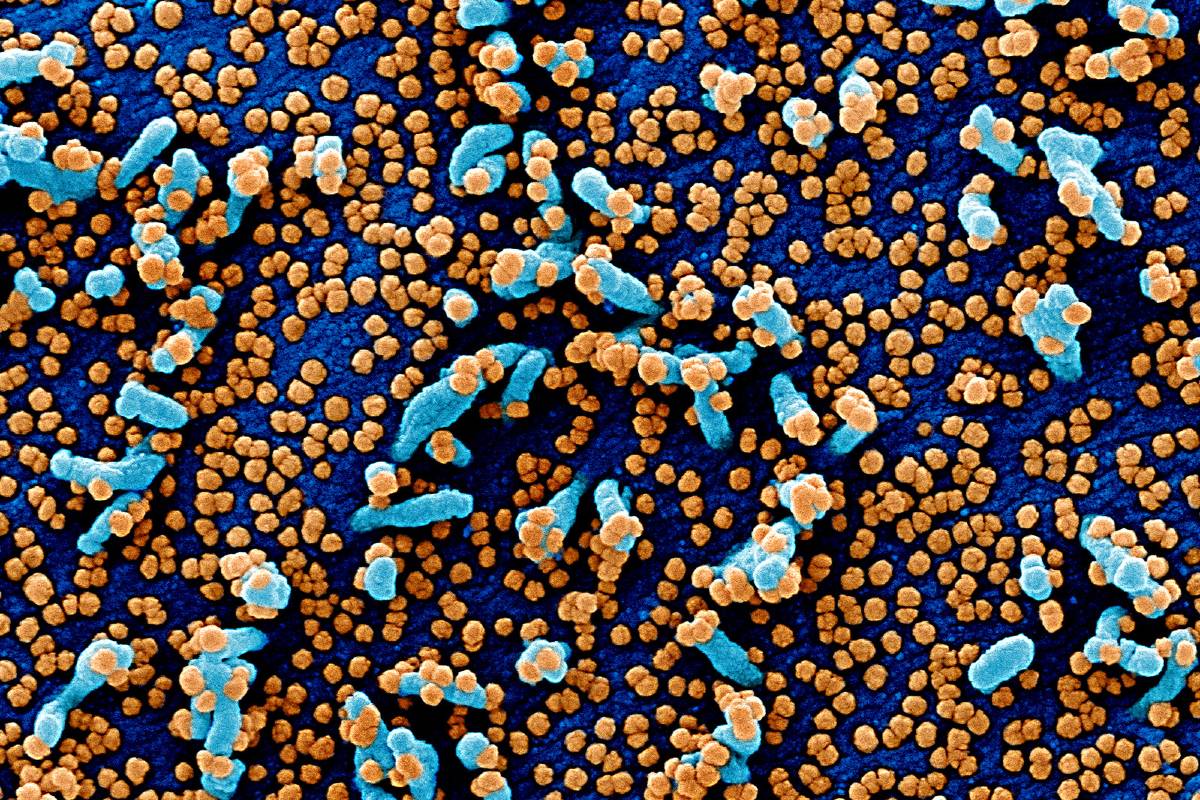
SARS-CoV-2 has a diameter of 60 nm to 140 nm and distinctive spikes, ranging from 9 nm to 12 nm, giving the virions the appearance of a solar corona (Figure 2).4 Through genetic recombination and variation, coronaviruses can adapt to and infect new hosts. Bats are thought to be a natural reservoir for SARS-CoV-2, but it has been suggested that humans became infected with SARS-CoV-2 via an intermediate host, such as the pangolin.5,6


The Host Defense Against SARS-CoV-2


Early in infection, SARS-CoV-2 targets cells, such as nasal and bronchial epithelial cells and pneumocytes, through the viral structural spike (S) protein that binds to the angiotensin-converting enzyme 2 (ACE2) receptor7 (Figure 2). The type 2 transmembrane serine protease (TMPRSS2), present in the host cell, promotes viral uptake by cleaving ACE2 and activating the SARS-CoV-2 S protein, which mediates coronavirus entry into host cells.7 ACE2 and TMPRSS2 are expressed in host target cells, particularly alveolar epithelial type II cells.8,9 Similar to other respiratory viral diseases, such as influenza, profound lymphopenia may occur in individuals with COVID-19 when SARS-CoV-2 infects and kills T lymphocyte cells. In addition, the viral inflammatory response, consisting of both the innate and the adaptive immune response (comprising humoral and cell-mediated immunity), impairs lymphopoiesis and increases lymphocyte apoptosis. Although upregulation of ACE2 receptors from ACE inhibitor and angiotensin receptor blocker medications has been hypothesized to increase susceptibility to SARS-CoV-2 infection, large observational cohorts have not found an association between these medications and risk of infection or hospital mortality due to COVID-19.10,11 For example, in a study 4480 patients with COVID-19 from Denmark, previous treatment with ACE inhibitors or angiotensin receptor blockers was not associated with mortality.11


In later stages of infection, when viral replication accelerates, epithelial-endothelial barrier integrity is compromised. In addition to epithelial cells, SARS-CoV-2 infects pulmonary capillary endothelial cells, accentuating the inflammatory response and triggering an influx of monocytes and neutrophils. Autopsy studies have shown diffuse thickening of the alveolar wall with mononuclear cells and macrophages infiltrating airspaces in addition to endothelialitis.12 Interstitial mononuclear inflammatory infiltrates and edema develop and appear as ground-glass opacities on computed tomographic imaging. Pulmonary edema filling the alveolar spaces with hyaline membrane formation follows, compatible with early-phase acute respiratory distress syndrome (ARDS).12 Bradykinin-dependent lung angioedema may contribute to disease.13 Collectively, endothelial barrier disruption, dysfunctional alveolar-capillary oxygen transmission, and impaired oxygen diffusion capacity are characteristic features of COVID-19.


In severe COVID-19, fulminant activation of coagulation and consumption of clotting factors occur.14,15 A report from Wuhan, China, indicated that 71% of 183 individuals who died of COVID-19 met criteria for diffuse intravascular coagulation.14 Inflamed lung tissues and pulmonary endothelial cells may result in microthrombi formation and contribute to the high incidence of thrombotic complications, such as deep venous thrombosis, pulmonary embolism, and thrombotic arterial complications (eg, limb ischemia, ischemic stroke, myocardial infarction) in critically ill patients.16 The development of viral sepsis, defined as life-threatening organ dysfunction caused by a dysregulated host response to infection, may further contribute to multiorgan failure.


Transmission of SARS-CoV-2 Infection


Epidemiologic data suggest that droplets expelled during face-to-face exposure during talking, coughing, or sneezing is the most common mode of transmission (Box 1). Prolonged exposure to an infected person (being within 6 feet for at least 15 minutes) and briefer exposures to individuals who are symptomatic (eg, coughing) are associated with higher risk for transmission, while brief exposures to asymptomatic contacts are less likely to result in transmission.25 Contact surface spread (touching a surface with virus on it) is another possible mode of transmission. Transmission may also occur via aerosols (smaller droplets that remain suspended in air), but it is unclear if this is a significant source of infection in humans outside of a laboratory setting.26,27 The existence of aerosols in physiological states (eg, coughing) or the detection of nucleic acid in the air does not mean that small airborne particles are infectious.28 Maternal COVID-19 is currently believed to be associated with low risk for vertical transmission. In most reported series, the mothers’ infection with SARS-CoV-2 occurred in the third trimester of pregnancy, with no maternal deaths and a favorable clinical course in the neonates.2931

Box 1.

Transmission, Symptoms, and Complications of Coronavirus Disease 2019 (COVID-19)

  • Transmission17 of severe acute respiratory syndrome coronavirus 2 (SARS-CoV-2) occurs primarily via respiratory droplets from face-to-face contact and, to a lesser degree, via contaminated surfaces. Aerosol spread may occur, but the role of aerosol spread in humans remains unclear. An estimated 48% to 62% of transmission may occur via presymptomatic carriers.

  • Common symptoms18 in hospitalized patients include fever (70%-90%), dry cough (60%-86%), shortness of breath (53%-80%), fatigue (38%), myalgias (15%-44%), nausea/vomiting or diarrhea (15%-39%), headache, weakness (25%), and rhinorrhea (7%). Anosmia or ageusia may be the sole presenting symptom in approximately 3% of individuals with COVID-19.

  • Common laboratory abnormalities19 among hospitalized patients include lymphopenia (83%), elevated inflammatory markers (eg, erythrocyte sedimentation rate, C-reactive protein, ferritin, tumor necrosis factor-α, IL-1, IL-6), and abnormal coagulation parameters (eg, prolonged prothrombin time, thrombocytopenia, elevated D-dimer [46% of patients], low fibrinogen).

  • Common radiographic findings of individuals with COVID-19 include bilateral, lower-lobe predominate infiltrates on chest radiographic imaging and bilateral, peripheral, lower-lobe ground-glass opacities and/or consolidation on chest computed tomographic imaging.

  • Common complications18,2024 among hospitalized patients with COVID-19 include pneumonia (75%); acute respiratory distress syndrome (15%); acute liver injury, characterized by elevations in aspartate transaminase, alanine transaminase, and bilirubin (19%); cardiac injury, including troponin elevation (7%-17%), acute heart failure, dysrhythmias, and myocarditis; prothrombotic coagulopathy resulting in venous and arterial thromboembolic events (10%-25%); acute kidney injury (9%); neurologic manifestations, including impaired consciousness (8%) and acute cerebrovascular disease (3%); and shock (6%).

  • Rare complications among critically ill patients with COVID-19 include cytokine storm and macrophage activation syndrome (ie, secondary hemophagocytic lymphohistiocytosis).


The clinical significance of SARS-CoV-2 transmission from inanimate surfaces is difficult to interpret without knowing the minimum dose of virus particles that can initiate infection. Viral load appears to persist at higher levels on impermeable surfaces, such as stainless steel and plastic, than permeable surfaces, such as cardboard.32 Virus has been identified on impermeable surfaces for up to 3 to 4 days after inoculation.32 Widespread viral contamination of hospital rooms has been documented.28 However, it is thought that the amount of virus detected on surfaces decays rapidly within 48 to 72 hours.32 Although the detection of virus on surfaces reinforces the potential for transmission via fomites (objects such as a doorknob, cutlery, or clothing that may be contaminated with SARS-CoV-2) and the need for adequate environmental hygiene, droplet spread via face-to-face contact remains the primary mode of transmission.


Viral load in the upper respiratory tract appears to peak around the time of symptom onset and viral shedding begins approximately 2 to 3 days prior to the onset of symptoms.33 Asymptomatic and presymptomatic carriers can transmit SARS-CoV-2.34,35 In Singapore, presymptomatic transmission has been described in clusters of patients with close contact (eg, through churchgoing or singing class) approximately 1 to 3 days before the source patient developed symptoms.34 Presymptomatic transmission is thought to be a major contributor to the spread of SARS-CoV-2. Modeling studies from China and Singapore estimated the percentage of infections transmitted from a presymptomatic individual as 48% to 62%.17 Pharyngeal shedding is high during the first week of infection at a time in which symptoms are still mild, which might explain the efficient transmission of SARS-CoV-2, because infected individuals can be infectious before they realize they are ill.36 Although studies have described rates of asymptomatic infection, ranging from 4% to 32%, it is unclear whether these reports represent truly asymptomatic infection by individuals who never develop symptoms, transmission by individuals with very mild symptoms, or transmission by individuals who are asymptomatic at the time of transmission but subsequently develop symptoms.3739 A systematic review on this topic suggested that true asymptomatic infection is probably uncommon.38


Although viral nucleic acid can be detectable in throat swabs for up to 6 weeks after the onset of illness, several studies suggest that viral cultures are generally negative for SARS-CoV-2 8 days after symptom onset.33,36,40 This is supported by epidemiological studies that have shown that transmission did not occur to contacts whose exposure to the index case started more than 5 days after the onset of symptoms in the index case.41 This suggests that individuals can be released from isolation based on clinical improvement. The Centers for Disease Control and Prevention recommend isolating for at least 10 days after symptom onset and 3 days after improvement of symptoms.42 However, there remains uncertainty about whether serial testing is required for specific subgroups, such as immunosuppressed patients or critically ill patients for whom symptom resolution may be delayed or older adults residing in short- or long-term care facilities.


Clinical Presentation


The mean (interquartile range) incubation period (the time from exposure to symptom onset) for COVID-19 is approximately 5 (2-7) days.43,44 Approximately 97.5% of individuals who develop symptoms will do so within 11.5 days of infection.43 The median (interquartile range) interval from symptom onset to hospital admission is 7 (3-9) days.45 The median age of hospitalized patients varies between 47 and 73 years, with most cohorts having a male preponderance of approximately 60%.44,46,47 Among patients hospitalized with COVID-19, 74% to 86% are aged at least 50 years.45,47


COVID-19 has various clinical manifestations (Box 1 and Box 2). In a study of 44 672 patients with COVID-19 in China, 81% of patients had mild manifestations, 14% had severe manifestations, and 5% had critical manifestations (defined by respiratory failure, septic shock, and/or multiple organ dysfunction).48 A study of 20 133 individuals hospitalized with COVID-19 in the UK reported that 17.1% were admitted to high-dependency or intensive care units (ICUs).47

Box 2.

Commonly Asked Questions About Coronavirus Disease 2019 (COVID-19)

  • How is severe acute respiratory syndrome coronavirus 2 (SARS-CoV-2) most commonly transmitted?

    • SARS-CoV-2 is most commonly spread via respiratory droplets (eg, from coughing, sneezing, shouting) during face-to-face exposure or by surface contamination.

  • What are the most common symptoms of COVID-19?

    • The 3 most common symptoms are fever, cough, and shortness of breath. Additional symptoms include weakness, fatigue, nausea, vomiting, diarrhea, changes to taste and smell.

  • How is the diagnosis made?

    • Diagnosis of COVID-19 is typically made by polymerase chain reaction testing of a nasopharyngeal swab. However, given the possibility of false-negative test results, clinical, laboratory, and imaging findings may also be used to make a presumptive diagnosis for individuals for whom there is a high index of clinical suspicion of infection.

  • What are current evidence-based treatments for individuals with COVID-19?

    • Supportive care, including supplemental oxygen, is the main treatment for most patients. Recent trials indicate that dexamethasone decreases mortality (subgroup analysis suggests benefit is limited to patients who require supplemental oxygen and who have symptoms for >7 d) and remdesivir improves time to recovery (subgroup analysis suggests benefit is limited to patients not receiving mechanical ventilation).

  • What percentage of people are asymptomatic carriers, and how important are they in transmitting the disease?

    • True asymptomatic infection is believed to be uncommon. The average time from exposure to symptoms onset is 5 days, and up to 62% of transmission may occur prior to the onset of symptoms.

  • Are masks effective at preventing spread?

    • Yes. Face masks reduce the spread of viral respiratory infection. N95 respirators and surgical masks both provide substantial protection (compared with no mask), and surgical masks provide greater protection than cloth masks. However, physical distancing is also associated with substantial reduction of viral transmission, with greater distances providing greater protection. Additional measures such as hand and environmental disinfection are also important.


Although only approximately 25% of infected patients have comorbidities, 60% to 90% of hospitalized infected patients have comorbidities.4549 The most common comorbidities in hospitalized patients include hypertension (present in 48%-57% of patients), diabetes (17%-34%), cardiovascular disease (21%-28%), chronic pulmonary disease (4%-10%), chronic kidney disease (3%-13%), malignancy (6%-8%), and chronic liver disease (<5%).45,46,49


The most common symptoms in hospitalized patients are fever (up to 90% of patients), dry cough (60%-86%), shortness of breath (53%-80%), fatigue (38%), nausea/vomiting or diarrhea (15%-39%), and myalgia (15%-44%).18,4447,49,50 Patients can also present with nonclassical symptoms, such as isolated gastrointestinal symptoms.18 Olfactory and/or gustatory dysfunctions have been reported in 64% to 80% of patients.5153 Anosmia or ageusia may be the sole presenting symptom in approximately 3% of patients.53


Complications of COVID-19 include impaired function of the heart, brain, lung, liver, kidney, and coagulation system. COVID-19 can lead to myocarditis, cardiomyopathy, ventricular arrhythmias, and hemodynamic instability.20,54 Acute cerebrovascular disease and encephalitis are observed with severe illness (in up to 8% of patients).21,52 Venous and arterial thromboembolic events occur in 10% to 25% in hospitalized patients with COVID-19.19,22 In the ICU, venous and arterial thromboembolic events may occur in up to 31% to 59% of patients with COVID-19.16,22


Approximately 17% to 35% of hospitalized patients with COVID-19 are treated in an ICU, most commonly due to hypoxemic respiratory failure. Among patients in the ICU with COVID-19, 29% to 91% require invasive mechanical ventilation.47,49,55,56 In addition to respiratory failure, hospitalized patients may develop acute kidney injury (9%), liver dysfunction (19%), bleeding and coagulation dysfunction (10%-25%), and septic shock (6%).18,19,23,49,56


Approximately 2% to 5% of individuals with laboratory-confirmed COVID-19 are younger than 18 years, with a median age of 11 years. Children with COVID-19 have milder symptoms that are predominantly limited to the upper respiratory tract, and rarely require hospitalization. It is unclear why children are less susceptible to COVID-19. Potential explanations include that children have less robust immune responses (ie, no cytokine storm), partial immunity from other viral exposures, and lower rates of exposure to SARS-CoV-2. Although most pediatric cases are mild, a small percentage (<7%) of children admitted to the hospital for COVID-19 develop severe disease requiring mechanical ventilation.57 A rare multisystem inflammatory syndrome similar to Kawasaki disease has recently been described in children in Europe and North America with SARS-CoV-2 infection.58,59 This multisystem inflammatory syndrome in children is uncommon (2 in 100 000 persons aged <21 years).60


Assessment and Diagnosis


Diagnosis of COVID-19 is typically made using polymerase chain reaction testing via nasal swab (Box 2). However, because of false-negative test result rates of SARS-CoV-2 PCR testing of nasal swabs, clinical, laboratory, and imaging findings may also be used to make a presumptive diagnosis.


Diagnostic Testing: Polymerase Chain Reaction and Serology


Reverse transcription polymerase chain reaction–based SARS-CoV-2 RNA detection from respiratory samples (eg, nasopharynx) is the standard for diagnosis. However, the sensitivity of testing varies with timing of testing relative to exposure. One modeling study estimated sensitivity at 33% 4 days after exposure, 62% on the day of symptom onset, and 80% 3 days after symptom onset.6163 Factors contributing to false-negative test results include the adequacy of the specimen collection technique, time from exposure, and specimen source. Lower respiratory samples, such as bronchoalveolar lavage fluid, are more sensitive than upper respiratory samples. Among 1070 specimens collected from 205 patients with COVID-19 in China, bronchoalveolar lavage fluid specimens had the highest positive rates of SARS-CoV-2 PCR testing results (93%), followed by sputum (72%), nasal swabs (63%), and pharyngeal swabs (32%).61 SARS-CoV-2 can also be detected in feces, but not in urine.61 Saliva may be an alternative specimen source that requires less personal protective equipment and fewer swabs, but requires further validation.64


Several serological tests can also aid in the diagnosis and measurement of responses to novel vaccines.62,65,66 However, the presence of antibodies may not confer immunity because not all antibodies produced in response to infection are neutralizing. Whether and how frequently second infections with SARS-CoV-2 occur remain unknown. Whether presence of antibody changes susceptibility to subsequent infection or how long antibody protection lasts are unknown. IgM antibodies are detectable within 5 days of infection, with higher IgM levels during weeks 2 to 3 of illness, while an IgG response is first seen approximately 14 days after symptom onset.62,65 Higher antibody titers occur with more severe disease.66 Available serological assays include point-of-care assays and high throughput enzyme immunoassays. However, test performance, accuracy, and validity are variable.67


Laboratory Findings


A systematic review of 19 studies of 2874 patients who were mostly from China (mean age, 52 years), of whom 88% were hospitalized, reported the typical range of laboratory abnormalities seen in COVID-19, including elevated serum C-reactive protein (increased in >60% of patients), lactate dehydrogenase (increased in approximately 50%-60%), alanine aminotransferase (elevated in approximately 25%), and aspartate aminotransferase (approximately 33%).24 Approximately 75% of patients had low albumin.24 The most common hematological abnormality is lymphopenia (absolute lymphocyte count <1.0 × 109/L), which is present in up to 83% of hospitalized patients with COVID-19.44,50 In conjunction with coagulopathy, modest prolongation of prothrombin times (prolonged in >5% of patients), mild thrombocytopenia (present in approximately 30% of patients) and elevated D-dimer values (present in 43%-60% of patients) are common.14,15,19,44,68 However, most of these laboratory characteristics are nonspecific and are common in pneumonia. More severe laboratory abnormalities have been associated with more severe infection.44,50,69 D-dimer and, to a lesser extent, lymphopenia seem to have the largest prognostic associations.69


Imaging


The characteristic chest computed tomographic imaging abnormalities for COVID-19 are diffuse, peripheral ground-glass opacities (Figure 3).70 Ground-glass opacities have ill-defined margins, air bronchograms, smooth or irregular interlobular or septal thickening, and thickening of the adjacent pleura.70 Early in the disease, chest computed tomographic imaging findings in approximately 15% of individuals and chest radiograph findings in approximately 40% of individuals can be normal.44 Rapid evolution of abnormalities can occur in the first 2 weeks after symptom onset, after which they subside gradually.70,71


Chest computed tomographic imaging findings are nonspecific and overlap with other infections, so the diagnostic value of chest computed tomographic imaging for COVID-19 is limited. Some patients admitted to the hospital with polymerase chain reaction testing–confirmed SARS-CoV-2 infection have normal computed tomographic imaging findings, while abnormal chest computed tomographic imaging findings compatible with COVID-19 occur days before detection of SARS-CoV-2 RNA in other patients.70,71


Treatment


Supportive Care and Respiratory Support


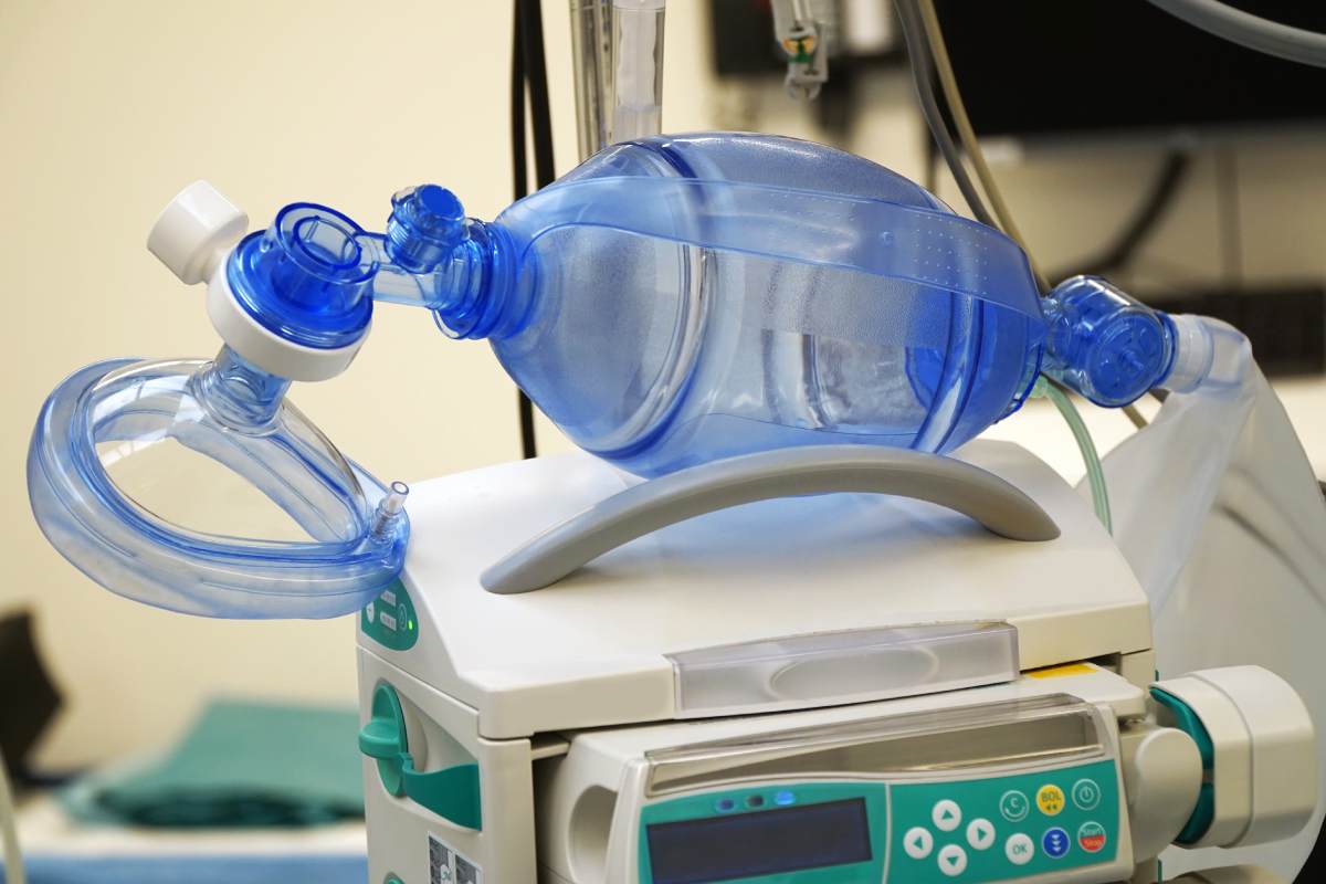
Currently, best practices for supportive management of acute hypoxic respiratory failure and ARDS should be followed.7274 Evidence-based guideline initiatives have been established by many countries and professional societies,7274 including guidelines that are updated regularly by the National Institutes of Health.74


More than 75% of patients hospitalized with COVID-19 require supplemental oxygen therapy. For patients who are unresponsive to conventional oxygen therapy, heated high-flow nasal canula oxygen may be administered.72 For patients requiring invasive mechanical ventilation, lung-protective ventilation with low tidal volumes (4-8 mL/kg, predicted body weight) and plateau pressure less than 30 mg Hg is recommended.72 Additionally, prone positioning, a higher positive end-expiratory pressure strategy, and short-term neuromuscular blockade with cisatracurium or other muscle relaxants may facilitate oxygenation. Although some patients with COVID-19–related respiratory failure have high lung compliance,75 they are still likely to benefit from lung-protective ventilation.76 Cohorts of patients with ARDS have displayed similar heterogeneity in lung compliance, and even patients with greater compliance have shown benefit from lower tidal volume strategies.76


The threshold for intubation in COVID-19–related respiratory failure is controversial, because many patients have normal work of breathing but severe hypoxemia.77 “Earlier” intubation allows time for a more controlled intubation process, which is important given the logistical challenges of moving patients to an airborne isolation room and donning personal protective equipment prior to intubation. However, hypoxemia in the absence of respiratory distress is well tolerated, and patients may do well without mechanical ventilation. Earlier intubation thresholds may result in treating some patients with mechanical ventilation unnecessarily and exposing them to additional complications. Currently, insufficient evidence exists to make recommendations regarding earlier vs later intubation.


In observational studies, approximately 8% of hospitalized patients with COVID-19 experience a bacterial or fungal co-infection, but up to 72% are treated with broad-spectrum antibiotics.78 Awaiting further data, it may be prudent to withhold antibacterial drugs in patients with COVID-19 and reserve them for those who present with radiological findings and/or inflammatory markers compatible with co-infection or who are immunocompromised and/or critically ill.72


Targeting the Virus and the Host Response


The following classes of drugs are being evaluated or developed for the management of COVID-19: antivirals (eg, remdesivir, favipiravir), antibodies (eg, convalescent plasma, hyperimmune immunoglobulins), anti-inflammatory agents (dexamethasone, statins), targeted immunomodulatory therapies (eg, tocilizumab, sarilumab, anakinra, ruxolitinib), anticoagulants (eg, heparin), and antifibrotics (eg, tyrosine kinase inhibitors). It is likely that different treatment modalities might have different efficacies at different stages of illness and in different manifestations of disease. Viral inhibition would be expected to be most effective early in infection, while, in hospitalized patients, immunomodulatory agents may be useful to prevent disease progression and anticoagulants may be useful to prevent thromboembolic complications.


More than 200 trials of chloroquine/hydroxychloroquine, compounds that inhibit viral entry and endocytosis of SARS-CoV-2 in vitro and may have beneficial immunomodulatory effects in vivo,79,80 have been initiated, but early data from clinical trials in hospitalized patients with COVID-19 have not demonstrated clear benefit.8183 A clinical trial of 150 patients in China admitted to the hospital for mild to moderate COVID-19 did not find an effect on negative conversion of SARS-CoV-2 by 28 days (the main outcome measure) when compared with standard of care alone.83 Two retrospective studies found no effect of hydroxychloroquine on risk of intubation or mortality among patients hospitalized for COVID-19.84,85 One of these retrospective multicenter cohort studies compared in-hospital mortality between those treated with hydroxychloroquine plus azithromycin (735 patients), hydroxychloroquine alone (271 patients), azithromycin alone (211 patients), and neither drug (221 patients), but reported no differences across the groups.84 Adverse effects are common, most notably QT prolongation with an increased risk of cardiac complications in an already vulnerable population.82,84 These findings do not support off-label use of (hydroxy)chloroquine either with or without the coadministration of azithromycin. Randomized clinical trials are ongoing and should provide more guidance.


Most antiviral drugs undergoing clinical testing in patients with COVID-19 are repurposed antiviral agents originally developed against influenza, HIV, Ebola, or SARS/MERS.79,86 Use of the protease inhibitor lopinavir-ritonavir, which disrupts viral replication in vitro, did not show benefit when compared with standard care in a randomized, controlled, open-label trial of 199 hospitalized adult patients with severe COVID-19.87 Among the RNA-dependent RNA polymerase inhibitors, which halt SARS-CoV-2 replication, being evaluated, including ribavirin, favipiravir, and remdesivir, the latter seems to be the most promising.79,88 The first preliminary results of a double-blind, randomized, placebo-controlled trial of 1063 adults hospitalized with COVID-19 and evidence of lower respiratory tract involvement who were randomly assigned to receive intravenous remdesivir or placebo for up to 10 days demonstrated that patients randomized to receive remdesivir had a shorter time to recovery than patients in the placebo group (11 vs 15 days).88 A separate randomized, open-label trial among 397 hospitalized patients with COVID-19 who did not require mechanical ventilation reported that 5 days of treatment with remdesivir was not different than 10 days in terms of clinical status on day 14.89 The effect of remdesivir on survival remains unknown.


Treatment with plasma obtained from patients who have recovered from viral infections was first reported during the 1918 flu pandemic. A first report of 5 critically ill patients with COVID-19 treated with convalescent plasma containing neutralizing antibody showed improvement in clinical status among all participants, defined as a combination of changes of body temperature, Sequential Organ Failure Assessment score, partial pressure of oxygen/fraction of inspired oxygen, viral load, serum antibody titer, routine blood biochemical index, ARDS, and ventilatory and extracorporeal membrane oxygenation supports before and after convalescent plasma transfusion status.90 However, a subsequent multicenter, open-label, randomized clinical trial of 103 patients in China with severe COVID-19 found no statistical difference in time to clinical improvement within 28 days among patients randomized to receive convalescent plasma vs standard treatment alone (51.9% vs 43.1%).91 However, the trial was stopped early because of slowing enrollment, which limited the power to detect a clinically important difference. Alternative approaches being studied include the use of convalescent plasma-derived hyperimmune globulin and monoclonal antibodies targeting SARS-CoV-2.92,93


Alternative therapeutic strategies consist of modulating the inflammatory response in patients with COVID-19. Monoclonal antibodies directed against key inflammatory mediators, such as interferon gamma, interleukin 1, interleukin 6, and complement factor 5a, all target the overwhelming inflammatory response following SARS-CoV-2 infection with the goal of preventing organ damage.79,86,94 Of these, the interleukin 6 inhibitors tocilizumab and sarilumab are best studied, with more than a dozen randomized clinical trials underway.94 Tyrosine kinase inhibitors, such as imatinib, are studied for their potential to prevent pulmonary vascular leakage in individuals with COVID-19.


Studies of corticosteroids for viral pneumonia and ARDS have yielded mixed results.72,73 However, the Randomized Evaluation of COVID-19 Therapy (RECOVERY) trial, which randomized 2104 patients with COVID-19 to receive 6 mg daily of dexamethasone for up to 10 days and 4321 to receive usual care, found that dexamethasone reduced 28-day all-cause mortality (21.6% vs 24.6%; age-adjusted rate ratio, 0.83 [95% CI, 0.74-0.92]; P < .001).95 The benefit was greatest in patients with symptoms for more than 7 days and patients who required mechanical ventilation. By contrast, there was no benefit (and possibility for harm) among patients with shorter symptom duration and no supplemental oxygen requirement. A retrospective cohort study of 201 patients in Wuhan, China, with confirmed COVID-19 pneumonia and ARDS reported that treatment with methylprednisolone was associated with reduced risk of death (hazard ratio, 0.38 [95% CI, 0.20-0.72]).69


Thromboembolic prophylaxis with subcutaneous low molecular weight heparin is recommended for all hospitalized patients with COVID-19.15,19 Studies are ongoing to assess whether certain patients (ie, those with elevated D-dimer) benefit from therapeutic anticoagulation.


Disparities


A disproportionate percentage of COVID-19 hospitalizations and deaths occurs in lower-income and minority populations.45,96,97 In a report by the Centers for Disease Control and Prevention of 580 hospitalized patients for whom race data were available, 33% were Black and 45% were White, while 18% of residents in the surrounding community were Black and 59% were White.45 The disproportionate prevalence of COVID-19 among Black patients was separately reported in a retrospective cohort study of 3626 patients with COVID-19 from Louisiana, in which 77% of patients hospitalized with COVID-19 and 71% of patients who died of COVID-19 were Black, but Black individuals comprised only 31% of the area population.97,98 This disproportionate burden may be a reflection of disparities in housing, transportation, employment, and health. Minority populations are more likely to live in densely populated communities or housing, depend on public transportation, or work in jobs for which telework was not possible (eg, bus driver, food service worker). Black individuals also have a higher prevalence of chronic health conditions than White individuals.98,99


Prognosis


Overall hospital mortality from COVID-19 is approximately 15% to 20%, but up to 40% among patients requiring ICU admission. However, mortality rates vary across cohorts, reflecting differences in the completeness of testing and case identification, variable thresholds for hospitalization, and differences in outcomes. Hospital mortality ranges from less than 5% among patients younger than 40 years to 35% for patients aged 70 to 79 years and greater than 60% for patients aged 80 to 89 years.46 Estimated overall death rates by age group per 1000 confirmed cases are provided in the Table. Because not all people who die during the pandemic are tested for COVID-19, actual numbers of deaths from COVID-19 are higher than reported numbers.


Although long-term outcomes from COVID-19 are currently unknown, patients with severe illness are likely to suffer substantial sequelae. Survival from sepsis is associated with increased risk for mortality for at least 2 years, new physical disability, new cognitive impairment, and increased vulnerability to recurrent infection and further health deterioration. Similar sequalae are likely to be seen in survivors of severe COVID-19.100


Prevention and Vaccine Development


COVID-19 is a potentially preventable disease. The relationship between the intensity of public health action and the control of transmission is clear from the epidemiology of infection around the world.25,101,102 However, because most countries have implemented multiple infection control measures, it is difficult to determine the relative benefit of each.103,104 This question is increasingly important because continued interventions will be required until effective vaccines or treatments become available. In general, these interventions can be divided into those consisting of personal actions (eg, physical distancing, personal hygiene, and use of protective equipment), case and contact identification (eg, test-trace-track-isolate, reactive school or workplace closure), regulatory actions (eg, governmental limits on sizes of gatherings or business capacity; stay-at-home orders; proactive school, workplace, and public transport closure or restriction; cordon sanitaire or internal border closures), and international border measures (eg, border closure or enforced quarantine). A key priority is to identify the combination of measures that minimizes societal and economic disruption while adequately controlling infection. Optimal measures may vary between countries based on resource limitations, geography (eg, island nations and international border measures), population, and political factors (eg, health literacy, trust in government, cultural and linguistic diversity).


The evidence underlying these public health interventions has not changed since the 1918 flu pandemic.105 Mathematical modeling studies and empirical evidence support that public health interventions, including home quarantine after infection, restricting mass gatherings, travel restrictions, and social distancing, are associated with reduced rates of transmission.101,102,106 Risk of resurgence follows when these interventions are lifted.


A human vaccine is currently not available for SARS-CoV-2, but approximately 120 candidates are under development. Approaches include the use of nucleic acids (DNA or RNA), inactivated or live attenuated virus, viral vectors, and recombinant proteins or virus particles.107,108 Challenges to developing an effective vaccine consist of technical barriers (eg, whether S or receptor-binding domain proteins provoke more protective antibodies, prior exposure to adenovirus serotype 5 [which impairs immunogenicity in the viral vector vaccine], need for adjuvant), feasibility of large-scale production and regulation (eg, ensuring safety and effectiveness), and legal barriers (eg, technology transfer and licensure agreements). The SARS-CoV-2 S protein appears to be a promising immunogen for protection, but whether targeting the full-length protein or only the receptor-binding domain is sufficient to prevent transmission remains unclear.108 Other considerations include the potential duration of immunity and thus the number of vaccine doses needed to confer immunity.62,108 More than a dozen candidate SARS-CoV-2 vaccines are currently being tested in phase 1-3 trials.


Other approaches to prevention are likely to emerge in the coming months, including monoclonal antibodies, hyperimmune globulin, and convalscent titer. If proved effective, these approaches could be used in high-risk individuals, including health care workers, other essential workers, and older adults (particularly those in nursing homes or long-term care facilities).


Limitations


This review has several limitations. First, information regarding SARS CoV-2 is limited. Second, information provided here is based on current evidence, but may be modified as more information becomes available. Third, few randomized trials have been published to guide management of COVID-19.


Conclusions


As of July 1, 2020, more than 10 million people worldwide had been infected with SARS-CoV-2. Many aspects of transmission, infection, and treatment remain unclear. Advances in prevention and effective management of COVID-19 will require basic and clinical investigation and public health and clinical interventions.


Section Editors: Edward Livingston, MD, Deputy Editor, and Mary McGrae McDermott, MD, Deputy Editor.


Submissions: We encourage authors to submit papers for consideration as a Review. Please contact Edward Livingston, MD, at Edward.livingston@jamanetwork.org or Mary McGrae McDermott, MD, at mdm608@northwestern.edu.


Back to top

Article Information

Accepted for Publication: June 30, 2020.

Corresponding Author: W. Joost Wiersinga, MD, PhD, Division of Infectious Diseases, Department of Medicine, Amsterdam UMC, location AMC, Meibergdreef 9, 1105 AZ Amsterdam, the Netherlands (w.j.wiersinga@amsterdamumc.nl).

Published Online: July 10, 2020. doi:10.1001/jama.2020.12839

Conflict of Interest Disclosures: Dr Wiersinga is supported by the Netherlands Organisation of Scientific Research outside the submitted work. Dr Prescott reported receiving grants from the US Agency for Healthcare Research and Quality (HCP by R01 HS026725), the National Institutes of Health/National Institute of General Medical Sciences, and the US Department of Veterans Affairs outside the submitted work, being the sepsis physician lead for the Hospital Medicine Safety Continuous Quality Initiative funded by BlueCross/BlueShield of Michigan, and serving on the steering committee for MI-COVID-19, a Michigan statewide registry to improve care for patients with COVID-19 in Michigan. Dr Rhodes reported being the co-chair of the Surviving Sepsis Campaign. Dr Cheng reported being a member of Australian government advisory committees, including those involved in COVID-19. No other disclosures were reported.

Disclaimer: This article does not represent the views of the US Department of Veterans Affairs or the US government. This material is the result of work supported with resources and use of facilities at the Ann Arbor VA Medical Center. The opinions in this article do not necessarily represent those of the Australian government or advisory committees.


References

1.

Zhu  N, Zhang  D, Wang  W,  et al; China Novel Coronavirus Investigating and Research Team.  A novel coronavirus from patients with pneumonia in China, 2019.   N Engl J Med. 2020;382(8):727-733. doi:10.1056/NEJMoa2001017PubMedGoogle ScholarCrossref

2.

Zhong  NS, Zheng  BJ, Li  YM,  et al.  Epidemiology and cause of severe acute respiratory syndrome (SARS) in Guangdong, People’s Republic of China, in February, 2003.   Lancet. 2003;362(9393):1353-1358. doi:10.1016/S0140-6736(03)14630-2PubMedGoogle ScholarCrossref

3.

Zaki  AM, van Boheemen  S, Bestebroer  TM, Osterhaus  AD, Fouchier  RA.  Isolation of a novel coronavirus from a man with pneumonia in Saudi Arabia.   N Engl J Med. 2012;367(19):1814-1820. doi:10.1056/NEJMoa1211721PubMedGoogle ScholarCrossref

4.

Goldsmith  CS, Tatti  KM, Ksiazek  TG,  et al.  Ultrastructural characterization of SARS coronavirus.   Emerg Infect Dis. 2004;10(2):320-326. doi:10.3201/eid1002.030913PubMedGoogle ScholarCrossref

5.

Lu  R, Zhao  X, Li  J,  et al.  Genomic characterisation and epidemiology of 2019 novel coronavirus: implications for virus origins and receptor binding.   Lancet. 2020;395(10224):565-574. doi:10.1016/S0140-6736(20)30251-8PubMedGoogle ScholarCrossref

6.

Lam  TT, Jia  N, Zhang  YW,  et al.  Identifying SARS-CoV-2-related coronaviruses in Malayan pangolins.   Nature. Published online March 26, 2020. doi:10.1038/s41586-020-2169-0PubMedGoogle Scholar

7.

Hoffmann  M, Kleine-Weber  H, Schroeder  S,  et al.  SARS-CoV-2 cell entry depends on ACE2 and TMPRSS2 and is blocked by a clinically proven protease inhibitor.   Cell. 2020;181(2):271-280. doi:10.1016/j.cell.2020.02.052PubMedGoogle Scholar

8.

Sungnak  W, Huang  N, Bécavin  C,  et al; HCA Lung Biological Network.  SARS-CoV-2 entry factors are highly expressed in nasal epithelial cells together with innate immune genes.   Nat Med. 2020;26(5):681-687. doi:10.1038/s41591-020-0868-6PubMedGoogle ScholarCrossref

9.

Zou  X, Chen  K, Zou  J, Han  P, Hao  J, Han  Z.  Single-cell RNA-seq data analysis on the receptor ACE2 expression reveals the potential risk of different human organs vulnerable to 2019-nCoV infection.   Front Med. 2020;14(2):185-192. doi:10.1007/s11684-020-0754-0PubMedGoogle ScholarCrossref

10.

Mancia  G, Rea  F, Ludergnani  M, Apolone  G, Corrao  G.  Renin-angiotensin-aldosterone system blockers and the risk of COVID-19.   N Engl J Med. 2020;382(25):2431-2440. doi:10.1056/NEJMoa2006923PubMedGoogle ScholarCrossref

11.

Fosbøl  EL, Butt  JH, Østergaard  L,  et al.  Association of angiotensin-converting enzyme inhibitor or angiotensin receptor blocker use with COVID-19 diagnosis and mortality.   JAMA. Published online June 19, 2020. doi:10.1001/jama.2020.11301
ArticlePubMedGoogle Scholar

12.

Xu  Z, Shi  L, Wang  Y,  et al.  Pathological findings of COVID-19 associated with acute respiratory distress syndrome.   Lancet Respir Med. 2020;8(4):420-422. doi:10.1016/S2213-2600(20)30076-XPubMedGoogle ScholarCrossref

13.

van de Veerdonk  FL, Netea  MG, van Deuren  M,  et al.  Kallikrein-kinin blockade in patients with COVID-19 to prevent acute respiratory distress syndrome.   Elife. Published online April 27, 2020. doi:10.7554/eLife.57555PubMedGoogle Scholar

14.

Tang  N, Li  D, Wang  X, Sun  Z.  Abnormal coagulation parameters are associated with poor prognosis in patients with novel coronavirus pneumonia.   J Thromb Haemost. 2020;18(4):844-847. doi:10.1111/jth.14768PubMedGoogle ScholarCrossref

15.

Thachil  J, Tang  N, Gando  S,  et al.  ISTH interim guidance on recognition and management of coagulopathy in COVID-19.   J Thromb Haemost. 2020;18(5):1023-1026. doi:10.1111/jth.14810PubMedGoogle ScholarCrossref

16.

Klok  FA, Kruip  MJHA, van der Meer  NJM,  et al.  Incidence of thrombotic complications in critically ill ICU patients with COVID-19.   Thromb Res. 2020;191:145-147. doi:10.1016/j.thromres.2020.04.013PubMedGoogle ScholarCrossref

17.

Ganyani  T, Kremer  C, Chen  D,  et al.  Estimating the generation interval for coronavirus disease (COVID-19) based on symptom onset data, March 2020.   Euro Surveill. 2020;25(17). doi:10.2807/1560-7917.ES.2020.25.17.2000257PubMedGoogle Scholar

18.

Mao  R, Qiu  Y, He  JS,  et al.  Manifestations and prognosis of gastrointestinal and liver involvement in patients with COVID-19: a systematic review and meta-analysis.   Lancet Gastroenterol Hepatol. 2020;5(7):667-678. doi:10.1016/S2468-1253(20)30126-6PubMedGoogle ScholarCrossref

19.

Levi  M, Thachil  J, Iba  T, Levy  JH.  Coagulation abnormalities and thrombosis in patients with COVID-19.   Lancet Haematol. 2020;7(6):e438-e440. doi:10.1016/S2352-3026(20)30145-9PubMedGoogle ScholarCrossref

20.

Long  B, Brady  WJ, Koyfman  A, Gottlieb  M.  Cardiovascular complications in COVID-19.   Am J Emerg Med. Published online April 18, 2020. doi:10.1016/j.ajem.2020.04.048PubMedGoogle Scholar

21.

Mao  L, Jin  H, Wang  M,  et al.  Neurologic manifestations of hospitalized patients with coronavirus disease 2019 in Wuhan, China.   JAMA Neurol. 2020;77(6):1-9. doi:10.1001/jamaneurol.2020.1127
ArticlePubMedGoogle ScholarCrossref

22.

Middeldorp  S, Coppens  M, van Haaps  TF,  et al.  Incidence of venous thromboembolism in hospitalized patients with COVID-19.   J Thromb Haemost. Published online May 5, 2020. doi:10.1111/jth.14888PubMedGoogle Scholar

23.

Chen  YT, Shao  SC, Hsu  CK, Wu  IW, Hung  MJ, Chen  YC.  Incidence of acute kidney injury in COVID-19 infection: a systematic review and meta-analysis.   Crit Care. 2020;24(1):346. doi:10.1186/s13054-020-03009-yPubMedGoogle ScholarCrossref

24.

Rodriguez-Morales  AJ, Cardona-Ospina  JA, Gutiérrez-Ocampo  E,  et al; Latin American Network of Coronavirus Disease 2019-COVID-19 Research (LANCOVID-19).  Clinical, laboratory and imaging features of COVID-19: a systematic review and meta-analysis.   Travel Med Infect Dis. 2020;34:101623. doi:10.1016/j.tmaid.2020.101623PubMedGoogle Scholar

25.

Chu  DK, Akl  EA, Duda  S,  et al; COVID-19 Systematic Urgent Review Group Effort (SURGE) study authors.  Physical distancing, face masks, and eye protection to prevent person-to-person transmission of SARS-CoV-2 and COVID-19: a systematic review and meta-analysis.   Lancet. 2020;395(10242):1973-1987. doi:10.1016/S0140-6736(20)31142-9PubMedGoogle ScholarCrossref

26.

Bourouiba  L.  Turbulent gas clouds and respiratory pathogen emissions: potential implications for reducing transmission of COVID-19.   JAMA. Published online March 26, 2020. doi:10.1001/jama.2020.4756
ArticlePubMedGoogle Scholar

27.

Lewis  D.  Is the coronavirus airborne? experts can’t agree.   Nature. 2020;580(7802):175. doi:10.1038/d41586-020-00974-wPubMedGoogle ScholarCrossref

28.

Chia  PY, Coleman  KK, Tan  YK,  et al; Singapore 2019 Novel Coronavirus Outbreak Research Team.  Detection of air and surface contamination by SARS-CoV-2 in hospital rooms of infected patients.   Nat Commun. 2020;11(1):2800. doi:10.1038/s41467-020-16670-2PubMedGoogle ScholarCrossref

29.

Dashraath  P, Wong  JLJ, Lim  MXK,  et al.  Coronavirus disease 2019 (COVID-19) pandemic and pregnancy.   Am J Obstet Gynecol. 2020;222(6):521-531. doi:10.1016/j.ajog.2020.03.021PubMedGoogle ScholarCrossref

30.

Chen  H, Guo  J, Wang  C,  et al.  Clinical characteristics and intrauterine vertical transmission potential of COVID-19 infection in nine pregnant women: a retrospective review of medical records.   Lancet. 2020;395(10226):809-815. doi:10.1016/S0140-6736(20)30360-3PubMedGoogle ScholarCrossref

31.

Zeng  L, Xia  S, Yuan  W,  et al.  Neonatal early-onset infection with SARS-CoV-2 in 33 neonates born to mothers with COVID-19 in Wuhan, China.   JAMA Pediatr. Published online March 26, 2020. doi:10.1001/jamapediatrics.2020.0878
ArticlePubMedGoogle Scholar

32.

van Doremalen  N, Bushmaker  T, Morris  DH,  et al.  Aerosol and surface stability of SARS-CoV-2 as compared with SARS-CoV-1.   N Engl J Med. 2020;382(16):1564-1567. doi:10.1056/NEJMc2004973PubMedGoogle ScholarCrossref

33.

He  X, Lau  EHY, Wu  P,  et al.  Temporal dynamics in viral shedding and transmissibility of COVID-19.   Nat Med. 2020;26(5):672-675. doi:10.1038/s41591-020-0869-5PubMedGoogle ScholarCrossref

34.

Wei  WE, Li  Z, Chiew  CJ, Yong  SE, Toh  MP, Lee  VJ.  Presymptomatic transmission of SARS-CoV-2—Singapore, January 23-March 16, 2020.   MMWR Morb Mortal Wkly Rep. 2020;69(14):411-415. doi:10.15585/mmwr.mm6914e1PubMedGoogle ScholarCrossref

35.

Bai  Y, Yao  L, Wei  T,  et al.  Presumed asymptomatic carrier transmission of COVID-19.   JAMA. 2020;323(14):1406-1407. doi:10.1001/jama.2020.2565
ArticlePubMedGoogle ScholarCrossref

36.

Wölfel  R, Corman  VM, Guggemos  W,  et al.  Virological assessment of hospitalized patients with COVID-2019.   Nature. 2020;581(7809):465-469. doi:10.1038/s41586-020-2196-xPubMedGoogle ScholarCrossref

37.

Park  SY, Kim  YM, Yi  S,  et al.  Coronavirus disease outbreak in call center, South Korea.   Emerg Infect Dis. 2020;26(8). doi:10.3201/eid2608.201274PubMedGoogle Scholar

38.

Byambasuren  O, Cardona  M, Bell  K, Clark  J, McLaws  M, Glasziou  P. Estimating the extent of asymptomatic COVID-19 and its potential for community transmission: systematic review and meta-analysis. MedRxiv. Preprint posted June 4, 2020. doi:10.1101/2020.05.10.20097543

39.

Tabata  S, Imai  K, Kawano  S,  et al.  Clinical characteristics of COVID-19 in 104 people with SARS-CoV-2 infection on the Diamond Princess cruise ship: a retrospective analysis.   Lancet Infect Dis. Published online June 12, 2020. doi:10.1016/S1473-3099(20)30482-5PubMedGoogle Scholar

40.

Sun  J, Xiao  J, Sun  R,  et al.  Prolonged persistence of SARS-CoV-2 RNA in body fluids.   Emerg Infect Dis. Published online May 8, 2020. doi:10.3201/eid2608.201097PubMedGoogle Scholar

41.

Cheng  HY, Jian  SW, Liu  DP,  et al.  Contact tracing assessment of COVID-19 transmission dynamics in Taiwan and risk at different exposure periods before and after symptom onset.   JAMA Intern Med. Published online May 1, 2020. doi:10.1001/jamainternmed.2020.2020
ArticlePubMedGoogle Scholar

42.

Symptom-based strategy to discontinue isolation for persons with COVID-19. Centers for Disease Control and Prevention website. Updated May 3, 2020. Accessed July 6, 2020. https://www.cdc.gov/coronavirus/2019-ncov/community/strategy-discontinue-isolation.html

43.

Lauer  SA, Grantz  KH, Bi  Q,  et al.  The incubation period of coronavirus disease 2019 (COVID-19) from publicly reported confirmed cases: estimation and application.   Ann Intern Med. 2020;172(9):577-582. doi:10.7326/M20-0504PubMedGoogle ScholarCrossref

44.

Guan  WJ, Ni  ZY, Hu  Y,  et al; China Medical Treatment Expert Group for Covid-19.  Clinical characteristics of coronavirus disease 2019 in China.   N Engl J Med. 2020;382(18):1708-1720. doi:10.1056/NEJMoa2002032PubMedGoogle ScholarCrossref

45.

Garg  S, Kim  L, Whitaker  M,  et al.  Hospitalization rates and characteristics of patients hospitalized with laboratory-confirmed coronavirus disease 2019—COVID-NET, 14 States, March 1-30, 2020.   MMWR Morb Mortal Wkly Rep. 2020;69(15):458-464. doi:10.15585/mmwr.mm6915e3PubMedGoogle ScholarCrossref

46.

Richardson  S, Hirsch  JS, Narasimhan  M,  et al; the Northwell COVID-19 Research Consortium.  Presenting characteristics, comorbidities, and outcomes among 5700 patients hospitalized with COVID-19 in the New York City area.   JAMA. 2020;323(20):2052-2059. doi:10.1001/jama.2020.6775
ArticlePubMedGoogle ScholarCrossref

47.

Docherty  AB, Harrison  EM, Green  CA,  et al; ISARIC4C investigators.  Features of 20 133 UK patients in hospital with COVID-19 using the ISARIC WHO Clinical Characterisation Protocol: prospective observational cohort study.   BMJ. 2020;369:m1985. doi:10.1136/bmj.m1985PubMedGoogle ScholarCrossref

48.

 The epidemiological characteristics of an outbreak of 2019 novel coronavirus diseases (COVID-19)—China, 2020.   China CDC Weekly. 2020;2:10.Google Scholar

49.

Grasselli  G, Zangrillo  A, Zanella  A,  et al; COVID-19 Lombardy ICU Network.  Baseline characteristics and outcomes of 1591 patients infected with SARS-CoV-2 admitted to ICUs of the Lombardy Region, Italy.   JAMA. 2020;323(16):1574-1581. doi:10.1001/jama.2020.5394
ArticlePubMedGoogle ScholarCrossref

50.

Huang  C, Wang  Y, Li  X,  et al.  Clinical features of patients infected with 2019 novel coronavirus in Wuhan, China.   Lancet. 2020;395(10223):497-506. doi:10.1016/S0140-6736(20)30183-5PubMedGoogle ScholarCrossref

51.

Lechien  JR, Chiesa-Estomba  CM, De Siati  DR,  et al.  Olfactory and gustatory dysfunctions as a clinical presentation of mild-to-moderate forms of the coronavirus disease (COVID-19): a multicenter European study.   Eur Arch Otorhinolaryngol. 2020;277(8):2251-2261. doi:10.1007/s00405-020-05965-1PubMedGoogle ScholarCrossref

52.

Helms  J, Kremer  S, Merdji  H,  et al.  Neurologic features in severe SARS-CoV-2 infection.   N Engl J Med. 2020;382(23):2268-2270. doi:10.1056/NEJMc2008597PubMedGoogle ScholarCrossref

53.

Spinato  G, Fabbris  C, Polesel  J,  et al.  Alterations in smell or taste in mildly symptomatic outpatients with SARS-CoV-2 infection.   JAMA. 2020;323(20):2089-2209. doi:10.1001/jama.2020.6771
ArticlePubMedGoogle ScholarCrossref

54.

Hendren  NS, Drazner  MH, Bozkurt  B, Cooper  LT  Jr.  Description and proposed management of the acute COVID-19 cardiovascular syndrome.   Circulation. 2020;141(23):1903-1914. doi:10.1161/CIRCULATIONAHA.120.047349PubMedGoogle ScholarCrossref

55.

Myers  LC, Parodi  SM, Escobar  GJ, Liu  VX.  Characteristics of hospitalized adults with COVID-19 in an integrated health care system in California.   JAMA. 2020;323(21):2195-2198. doi:10.1001/jama.2020.7202
ArticlePubMedGoogle ScholarCrossref

56.

Yang  X, Yu  Y, Xu  J,  et al.  Clinical course and outcomes of critically ill patients with SARS-CoV-2 pneumonia in Wuhan, China: a single-centered, retrospective, observational study.   Lancet Respir Med. 2020;8(5):475-481. doi:10.1016/S2213-2600(20)30079-5PubMedGoogle ScholarCrossref

57.

Götzinger  F, Santiago-García  B, Noguera-Julián  A, Lanaspa  M, Lancella  L, Carducci  FIC.  COVID-19 in children and adolescents in Europe: a multinational, multicentre cohort study.   Lancet Child Adolesc Health. Published online June 25, 2020. doi:10.1016/S2352-4642(20)30177-2PubMedGoogle Scholar

58.

Verdoni  L, Mazza  A, Gervasoni  A,  et al.  An outbreak of severe Kawasaki-like disease at the Italian epicentre of the SARS-CoV-2 epidemic: an observational cohort study.   Lancet. 2020;395(10239):1771-1778. doi:10.1016/S0140-6736(20)31103-XPubMedGoogle ScholarCrossref

59.

Whittaker  E, Bamford  A, Kenny  J,  et al.  Clinical characteristics of 58 children with a pediatric inflammatory multisystem syndrome temporally associated with SARS-CoV-2.   JAMA. Published online June 8, 2020. doi:10.1001/jama.2020.10369
ArticlePubMedGoogle Scholar

60.

Levin  M.  Childhood multisystem inflammatory syndrome: a new challenge in the pandemic.   N Engl J Med. Published online June 29, 2020. doi:10.1056/NEJMe2023158PubMedGoogle Scholar

61.

Wang  W, Xu  Y, Gao  R,  et al.  Detection of SARS-CoV-2 in different types of clinical specimens.   JAMA. 2020;323(18):1843-1844. doi:10.1001/jama.2020.3786
ArticlePubMedGoogle Scholar

62.

Sethuraman  N, Jeremiah  SS, Ryo  A.  Interpreting diagnostic tests for SARS-CoV-2.   JAMA. Published online May 6, 2020. doi:10.1001/jama.2020.8259
ArticlePubMedGoogle Scholar

63.

Kucirka  LM, Lauer  SA, Laeyendecker  O, Boon  D, Lessler  J.  Variation in false-negative rate of reverse transcriptase polymerase chain reaction-based SARS-CoV-2 tests by time since exposure.   Ann Intern Med. Published online May 13, 2020. doi:10.7326/M20-1495PubMedGoogle Scholar

64.

Williams  E, Bond  K, Zhang  B, Putland  M, Williamson  DA.  Saliva as a non-invasive specimen for detection of SARS-CoV-2.   J Clin Microbiol. Published online April 21, 2020. doi:10.1128/JCM.00776-20PubMedGoogle Scholar

65.

Guo  L, Ren  L, Yang  S,  et al.  Profiling early humoral response to diagnose novel coronavirus disease (COVID-19).   Clin Infect Dis. Published online March 21, 2020. doi:10.1093/cid/ciaa310PubMedGoogle Scholar

66.

Zhao  J, Yuan  Q, Wang  H,  et al.  Antibody responses to SARS-CoV-2 in patients of novel coronavirus disease 2019.   Clin Infect Dis. Published online March 28, 2020. doi:10.1093/cid/ciaa344PubMedGoogle Scholar

67.

Bond  K, Nicholson  S, Hoang  T, Catton  M, Howden  B, Williamson  D. Post-Market Validation of Three Serological Assays for COVID-19. Office of Health Protection, Commonwealth Government of Australia; 2020.

68.

Chen  N, Zhou  M, Dong  X,  et al.  Epidemiological and clinical characteristics of 99 cases of 2019 novel coronavirus pneumonia in Wuhan, China: a descriptive study.   Lancet. 2020;395(10223):507-513. doi:10.1016/S0140-6736(20)30211-7PubMedGoogle ScholarCrossref

69.

Wu  C, Chen  X, Cai  Y,  et al.  Risk factors associated with acute respiratory distress syndrome and death in patients with coronavirus disease 2019 pneumonia in Wuhan, China.   JAMA Intern Med. Published online March 13, 2020. doi:10.1001/jamainternmed.2020.0994
ArticlePubMedGoogle Scholar

70.

Shi  H, Han  X, Jiang  N,  et al.  Radiological findings from 81 patients with COVID-19 pneumonia in Wuhan, China: a descriptive study.   Lancet Infect Dis. 2020;20(4):425-434. doi:10.1016/S1473-3099(20)30086-4PubMedGoogle ScholarCrossref

71.

Bernheim  A, Mei  X, Huang  M,  et al.  Chest CT findings in coronavirus disease-19 (COVID-19): relationship to duration of infection.   Radiology. 2020;295(3):200463. doi:10.1148/radiol.2020200463PubMedGoogle Scholar

72.

Alhazzani  W, Møller  MH, Arabi  YM,  et al.  Surviving Sepsis Campaign: guidelines on the management of critically ill adults with coronavirus disease 2019 (COVID-19).   Intensive Care Med. 2020;46(5):854-887. doi:10.1007/s00134-020-06022-5PubMedGoogle ScholarCrossref

73.

Wilson  KC, Chotirmall  SH, Bai  C, Rello  J; International Task Force on COVID-19. COVID-19: Interim Guidance on Management Pending Empirical Evidence. American Thoracic Society; 2020. Accessed July 7, 2020. https://www.thoracic.org/covid/covid-19-guidance.pdf

74.

Coronavirus disease 2019 (COVID-19) treatment guidelines. National Institutes of Health website. Updated June 25, 2020. Accessed July 1, 2020. https://www.covid19treatmentguidelines.nih.gov/

75.

Marini  JJ, Gattinoni  L.  Management of COVID-19 respiratory distress.   JAMA. Published online April 24, 2020. doi:10.1001/jama.2020.6825
ArticlePubMedGoogle Scholar

76.

Hager  DN, Krishnan  JA, Hayden  DL, Brower  RG; ARDS Clinical Trials Network.  Tidal volume reduction in patients with acute lung injury when plateau pressures are not high.   Am J Respir Crit Care Med. 2005;172(10):1241-1245. doi:10.1164/rccm.200501-048CPPubMedGoogle ScholarCrossref

77.

Tobin  MJ.  Basing respiratory management of COVID-19 on physiological principles.   Am J Respir Crit Care Med. 2020;201(11):1319-1320. doi:10.1164/rccm.202004-1076EDPubMedGoogle ScholarCrossref

78.

Rawson  TM, Moore  LSP, Zhu  N,  et al.  Bacterial and fungal co-infection in individuals with coronavirus: a rapid review to support COVID-19 antimicrobial prescribing.   Clin Infect Dis. Published online May 2, 2020. doi:10.1093/cid/ciaa530PubMedGoogle Scholar

79.

Sanders  JM, Monogue  ML, Jodlowski  TZ, Cutrell  JB.  Pharmacologic treatments for coronavirus disease 2019 (COVID-19).   JAMA. Published online April 13, 2020. doi:10.1001/jama.2020.6019
ArticlePubMedGoogle Scholar

80.

Wang  M, Cao  R, Zhang  L,  et al.  Remdesivir and chloroquine effectively inhibit the recently emerged novel coronavirus (2019-nCoV) in vitro.   Cell Res. 2020;30(3):269-271. doi:10.1038/s41422-020-0282-0PubMedGoogle ScholarCrossref

81.

Magagnoli  J, Narendran  S, Pereira  F,  et al. Outcomes of hydroxychloroquine usage in United States veterans hospitalized with COVID-19. MedRxiv. Preprint posted June 5, 2020. doi:10.1016/j.medj.2020.06.001

82.

Mahévas  M, Tran  VT, Roumier  M,  et al.  Clinical efficacy of hydroxychloroquine in patients with covid-19 pneumonia who require oxygen: observational comparative study using routine care data.   BMJ. 2020;369:m1844. doi:10.1136/bmj.m1844PubMedGoogle ScholarCrossref

83.

Tang  W, Cao  Z, Han  M,  et al.  Hydroxychloroquine in patients with mainly mild to moderate coronavirus disease 2019: open label, randomised controlled trial.   BMJ. 2020;369:m1849. doi:10.1136/bmj.m1849PubMedGoogle ScholarCrossref

84.

Rosenberg  ES, Dufort  EM, Udo  T,  et al.  Association of treatment with hydroxychloroquine or azithromycin with in-hospital mortality in patients with COVID-19 in New York State.   JAMA. 2020;323(24):2493-2502. doi:10.1001/jama.2020.8630
ArticlePubMedGoogle ScholarCrossref

85.

Geleris  J, Sun  Y, Platt  J,  et al.  Observational study of hydroxychloroquine in hospitalized patients with COVID-19.   N Engl J Med. 2020;382(25):2411-2418. doi:10.1056/NEJMoa2012410PubMedGoogle ScholarCrossref

86.

Scavone  C, Brusco  S, Bertini  M,  et al.  Current pharmacological treatments for COVID-19: what’s next?   Br J Pharmacol. Published online April 24, 2020. doi:10.1111/bph.15072PubMedGoogle Scholar

87.

Cao  B, Wang  Y, Wen  D,  et al.  A trial of lopinavir-ritonavir in adults hospitalized with severe Covid-19.   N Engl J Med. 2020;382(19):1787-1799. doi:10.1056/NEJMoa2001282PubMedGoogle ScholarCrossref

88.

Beigel  JH, Tomashek  KM, Dodd  LE,  et al.  Remdesivir for the treatment of COVID-19: preliminary report.   N Engl J Med. Published online May 22, 2020. doi:10.1056/NEJMoa2007764PubMedGoogle Scholar

89.

Goldman  JD, Lye  DCB, Hui  DS,  et al.  Remdesivir for 5 or 10 days in patients with severe COVID-19.   N Engl J Med. Published online May 27, 2020. doi:10.1056/NEJMoa2015301PubMedGoogle Scholar

90.

Shen  C, Wang  Z, Zhao  F,  et al.  Treatment of 5 critically ill patients with COVID-19 with convalescent plasma.   JAMA. 2020;323(16):1582-1589. doi:10.1001/jama.2020.4783
ArticlePubMedGoogle ScholarCrossref

91.

Li  L, Zhang  W, Hu  Y,  et al.  Effect of convalescent plasma therapy on time to clinical improvement in patients with severe and life-threatening COVID-19: a randomized clinical trial.   JAMA. Published online June 3, 2020. doi:10.1001/jama.2020.10044
ArticlePubMedGoogle Scholar

92.

Wang  C, Li  W, Drabek  D,  et al.  A human monoclonal antibody blocking SARS-CoV-2 infection.   Nat Commun. 2020;11(1):2251. doi:10.1038/s41467-020-16256-yPubMedGoogle ScholarCrossref

93.

Brouwer  PJM, Caniels  TG, van der Straten  K,  et al.  Potent neutralizing antibodies from COVID-19 patients define multiple targets of vulnerability.   Science. Published online June 15, 2020. doi:10.1126/science.abc5902.PubMedGoogle Scholar

94.

Alzghari  SK, Acuña  VS.  Supportive treatment with tocilizumab for COVID-19: a systematic review.   J Clin Virol. 2020;127:104380. doi:10.1016/j.jcv.2020.104380PubMedGoogle Scholar

95.

Horby  P, Lim  WS, Emberson  J, Mafham  M, Bell  J,  et al. Effect of dexamethasone in hospitalized patients with COVID-19: preliminary report. medRxiv. Published online June 22, 2020. doi:10.1101/2020.06.22.20137273:24

96.

Wadhera  RK, Wadhera  P, Gaba  P,  et al.  Variation in COVID-19 hospitalizations and deaths across New York City boroughs.   JAMA. 2020;323(21):2192-2195. doi:10.1001/jama.2020.7197
ArticlePubMedGoogle ScholarCrossref

97.

Price-Haywood  EG, Burton  J, Fort  D, Seoane  L.  Hospitalization and mortality among black patients and white patients with COVID-19.   N Engl J Med. 2020;382(26):2534-2543. doi:10.1056/NEJMsa2011686PubMedGoogle ScholarCrossref

98.

COVID-19 in racial and ethnic minority groups. Centers for Disease Control and Prevention website. Updated June 25, 2020. Accessed July 7, 2020. https://www.cdc.gov/coronavirus/2019-ncov/need-extra-precautions/racial-ethnic-minorities.html

99.

Pham  TM, Carpenter  JR, Morris  TP, Sharma  M, Petersen  I.  Ethnic differences in the prevalence of type 2 diabetes diagnoses in the UK: cross-sectional analysis of the health improvement network primary care database.   Clin Epidemiol. 2019;11:1081-1088. doi:10.2147/CLEP.S227621Google ScholarCrossref

100.

Prescott  HC, Angus  D.  Enhancing recovery from sepsis: a review.   JAMA. 2018;319(1):62-75. doi:10.1001/jama.2017.17687
ArticleGoogle ScholarCrossref

101.

Jüni  P, Rothenbühler  M, Bobos  P,  et al.  Impact of climate and public health interventions on the COVID-19 pandemic: a prospective cohort study.   CMAJ. 2020;192(21):E566-E573. doi:10.1503/cmaj.200920PubMedGoogle ScholarCrossref

102.

Pan  A, Liu  L, Wang  C,  et al.  Association of public health interventions with the epidemiology of the COVID-19 outbreak in Wuhan, China.   JAMA. 2020;323(19):1-9. doi:10.1001/jama.2020.6130
ArticlePubMedGoogle ScholarCrossref

103.

Coronavirus government response tracker. University of Oxford website. Accessed June 24, 2020. https://www.bsg.ox.ac.uk/research/research-projects/coronavirus-government-response-tracker

104.

Flaxman  S, Mishra  S, Gandy  A,  et al.  Estimating the effects of nonpharmaceutical interventions on COVID-19 in Europe.   Nature. Published online June 8, 2020. doi:10.1038/s41586-020-2405-7PubMedGoogle Scholar

105.

Markel  H, Lipman  HB, Navarro  JA,  et al.  Nonpharmaceutical interventions implemented by US cities during the 1918-1919 influenza pandemic.   JAMA. 2007;298(6):644-654. doi:10.1001/jama.298.6.644
ArticlePubMedGoogle ScholarCrossref

106.

Xiao  Y, Tang  B, Wu  J, Cheke  RA, Tang  S.  Linking key intervention timings to rapid decline of the COVID-19 effective reproductive number to quantify lessons from mainland China.   Int J Infect Dis. Published online June 11, 2020. doi:10.1016/j.ijid.2020.06.030PubMedGoogle Scholar

107.

Thanh Le  T, Andreadakis  Z, Kumar  A,  et al.  The COVID-19 vaccine development landscape.   Nat Rev Drug Discov. 2020;19(5):305-306. doi:10.1038/d41573-020-00073-5PubMedGoogle ScholarCrossref

108.

Lurie  N, Saville  M, Hatchett  R, Halton  J.  Developing Covid-19 vaccines at pandemic speed.   N Engl J Med. 2020;382(21):1969-1973. doi:10.1056/NEJMp2005630PubMedGoogle ScholarCrossref

#Face Mask Type Matters When #Sterilizing, Study Finds

Postado em Atualizado em


When sterilizing face masks, the type of face mask and the method of sterilization have a bearing on subsequent filtration efficiency, according to researchers. The greatest reduction in filtration efficiency after sterilization occurred with surgical face masks.

With plasma vapor hydrogen peroxide (H2O2) sterilization, filtration efficiency of N95 and KN95 masks was maintained at more than 95%, but for surgical face masks, filtration efficiency was reduced to less than 95%. With chlorine dioxide (ClO2) sterilization, on the other hand, filtration efficiency was maintained at above 95% for N95 masks, but for KN95 and surgical face masks, filtration efficiency was reduced to less than 80%.

In a research letter published online June 15 in JAMA Network Open, researchers from the University of Oklahoma Health Sciences Center, Oklahoma City, report the results of a study of the two sterilization techniques on the pressure drop and filtration efficiency of N95, KN95, and surgical face masks.

“The H2O2 treatment showed a small effect on the overall filtration efficiency of the tested masks, but the ClO2 treatment showed marked reduction in the overall filtration efficiency of the KN95s and surgical face masks. All pressure drop changes were within the acceptable range,” the researchers write.

The study did not evaluate the effect of repeated sterilizations on face masks.

Five masks of each type were sterilized with either H2Oor CIO2. Masks were then placed in a test chamber, and a salt aerosol was nebulized to assess both upstream and downstream filtration as well as pressure drop. The researchers used a mobility particle sizer to measure particle number concentration from 16.8 nm to 514 nm. An acceptable pressure drop was defined as a drop of less than 1.38 inches of water (35 mm) for inhalation.

Although pressure drop changes were within the acceptable range for all three mask types following sterlization with either method, H2Osterilization yielded the least reduction in filtration efficacy in all cases. After sterilization with H2O2, filtration efficiencies were 96.6%, 97.1%, and 91.6% for the N95s, KN95s, and the surgical face masks, respectively. In contrast, filtration efficiencies after ClO2 sterilization were 95.1%, 76.2%, and 77.9%, respectively.

The researchers note that although overall filtration efficiency was maintained with ClOsterilization, there was a significant drop in efficiency with respect to particles of approximately 300 nm (0.3 microns) in size. For particles of that size, mean filtration efficiency decreased to 86.2% for N95s, 40.8% for KN95s, and 47.1% for surgical face masks.

The testing described in the report is “quite affordable at $350 per mask type, so it is hard to imagine any healthcare provider cannot set aside a small budget to conduct such an important test,” author Evan Floyd, PhD, told Medscape Medical News.

Given the high demand for effective face masks and the current risk for counterfeit products, Floyd suggested that individual facilities test all masks intended for use by healthcare workers before and after sterilization procedures.

“However, if for some reason testing is not an option, we would recommend sticking to established brands and suppliers, perhaps reach out to your state health department or a local representative of the strategic stockpile of PPE,” he noted.

The authors acknowledge that further studies using a larger sample size and a greater variety of masks, as well as studies to evaluate different sterilization techniques, are required. Further, “measuring the respirator’s filtration efficiency by aerosol size instead of only measuring the overall filtration efficiency” should also be considered. Such an approach would enable researchers to evaluate the degree to which masks protect against specific infectious agents.


JAMA Netw Open. Published online June 15, 2020. Full text

#Covid-19: un fármaco “barato y de fácil acceso” reduce el riesgo de muerte en pacientes con #ventilación asistida

Postado em

Se trata de la dexametasona, un tratamiento a base de esteroides que, según los investigadores, es “un gran avance”

Este fármaco reduciría en un tercio el riesgo de muerte de aquellos pacientes que se encuentran enchufados a ventiladores.

Un fármaco barato y de fácil acceso en todo el mundo llamado dexametasona puede ayudar a salvar vidas de pacientes que se encuentran graves a causa del coronavirus, según un estudio de la Universidad de Oxford divulgado este martes.

El equipo investigador cree que el tratamiento a base de dosis bajas de esteroides supone un gran avance en la lucha contra la Covid-19, al reducir el riesgo de muerte en un tercio de aquellos pacientes que se encuentran enchufados a ventiladores.

En cuanto a los que precisan de una abordaje de la enfermedad con oxígeno, el citado fármaco reduce las muertes en una quinta parte, de acuerdo con estos hallazgos.

Esta medicina es una de las que se están empleando en el considerado mayor ensayo clínico del mundo, donde se experimenta con tratamientos existentes para otros males con el objetivo de ver si también funcionan para combatir el coronavirus.

5.000 vidas

Según estimaciones de los investigadores, si ese fármaco hubiera estado disponible en este país desde el principio de la pandemia, se habrían podido salvar hasta 5.000 vidas.

Además, debido a su bajo coste, consideran que podría ser muy beneficioso en los países pobres que afrontan grandes números de enfermos de COVID-19.

Aproximadamente 19 de cada 20 pacientes que se infectan de coronavirus mejoran sin tener que ser hospitalizados, recuerda el estudio.

De aquellos que han de ser ingresados en un centro médico, la mayoría también experimenta una mejoría, si bien algunos podrían necesitar oxígeno o ventilación mecánica. Estos últimos, según el estudio, son los considerados pacientes de alto riesgo a los que la dexametasona parece ayudar.

Ese fármaco se emplea ya para reducir inflamaciones en el caso de otras condiciones médicas y ayuda, al parecer, a detener parte del daño que se origina cuando el sistema inmunológico se sobreactiva mientras intenta luchar contra el coronavirus.

“Reduce la mortalidad”

En este ensayo clínico participaron unos 2.000 pacientes de hospitales, a los que se administró la medicina y su evolución se comparó con otros 4.000 enfermos a los que no se les prescribió.

Para aquellos pacientes conectados a ventiladores mecánicos, la dexametasona redujo el riesgo de muerte de un 40 a un 28%, al tiempo que en el caso de los enfermos que precisaron de oxígeno, el tratamiento redujo el riesgo mortal de un 25 a un 20%.

“Este es el único fármaco hasta la fecha que ha mostrado que reduce la mortalidad y la reduce de manera significativa. Es un gran avance“, afirmó el investigador principal del estudio, Peter Horby.

Para Martin Landray, otro de los científicos involucrados, los hallazgos sugieren que de cada ocho pacientes tratados que precisan de respiración asistida por ventiladores mecánicos, se podría salvar una vida.

En cuanto a los que necesitan abordaje con oxígeno, se salva una vida de cada 20-25, agregó.

“Hay un claro beneficio. El tratamiento consta de 10 días de dexametasona y cuesta unas 5 libras (5,5 euros/6,2 dólares) por paciente. Así que esencialmente cuesta 35 libras (38 euros/43 dólares) salvar una vida. Es un fármaco que está disponible en todo el globo”, remarcó Landray.

Según el experimento, la dexametasona no parece ayudar a personas que presentan síntomas leves de coronavirus -aquellos que no necesitan asistencia para respirar-.

El ensayo lleva funcionando desde el pasado marzo y en esas pruebas se ha incluido también el producto empleado para tratar la Malaria, la hidroxicloroquina, que ahora ha sido desechado ante el temor de que incremente el número de muertes y dé problemas coronarios.

Otro denominado remdesivir, un tratamiento antiviral que parece acortar el periodo de recuperación en pacientes con Covid-19, ya está disponible en el servicio público de salud de este país.