Mês: agosto 2019

#’Wasabi receptor’ may advance treatments for chronic pain

Postado em

Scorpion toxin targets the “wasabi receptor,” a specific receptor in nerve cells that helps humans react to wasabi, cigarette smoke, and environmental pollutants. According to new research, the toxin’s unusual mechanism of action means that it could help scientists learn more about chronic pain.
person seen from the back, having back pain

New research furthers scientists’ understanding of chronic pain.

Researchers from the University of California, San Francisco (UCSF) and the University of Queensland in Brisbane, Australia, conducted the study.

For the research, the scientists isolated a toxin called WaTx from the venom of the Australian Black Rock scorpion.

They discovered this toxin when they were researching animal venom compounds that could target the wasabi receptor, which is present throughout the human body in sensory nerve cells.

The findings of the study appear in the journal Cell.

How scorpion toxins affect nerves

The scientists’ goal in isolating compounds in venom was ultimately to study the wasabi receptor, a sensory receptor that is also called TRPA1.

Upon activation, this receptor opens up and allows ions to flow into the cell, which triggers pain and inflammation.

“Think of TRPA1 as the body’s ‘fire alarm’ for chemical irritants in the environment,” says John Lin King, a doctoral student in UCSF’s neuroscience graduate program and lead author of the study.

“When this receptor encounters a potentially harmful compound — specifically, a class of chemicals known as ‘reactive electrophiles,’ which can cause significant damage to cells — it is activated to let you know you’re being exposed to something dangerous that you need to remove yourself from.”

Other electrophile-containing substances that can trigger TRPA1 include cigarette smoke and environmental pollutants.

These irritants produce a response in the surface cells of the airways, which can lead to inflammation and cause coughing fits and breathing issues.

Chemicals in specific foods, such as wasabi, mustard, ginger, garlic, and onions, can also generate a response in nerve cells by targetting this receptor.

How the wasabi receptor toxin is different

Although the scorpion toxin triggers the wasabi receptor in the same way as these other substances, using the same sites on the receptor, it activates it in a different way. This mechanism was previously unknown.

The team found that WaTx contains a particular sequence of amino acids that enables it to pass directly through the membrane of a cell into its interior. This is an unusual feat, of which few other proteins are capable.

By forcing itself into the cell in this manner, WaTx bypasses the typical route that restricts what can enter.


MedicalNewsToday

#Symptoms, causes, and treatment of chronic kidney disease

Postado em Atualizado em

Resultado de imagem para chronic kidney disease

Chronic kidney disease is a slow and progressive loss of kidney function over a period of several years. Eventually, a person will develop permanent kidney failure.

Chronic kidney disease, also known as chronic renal failure, chronic renal disease, or chronic kidney failure, is much more widespread than people realize; it often goes undetected and undiagnosed until the disease is well advanced.

It is not unusual for people to realize they have chronic kidney failure only when their kidney function is down to 25 percent of normal.

As kidney failure advances and the organ’s function is severely impaired, dangerous levels of waste and fluid can rapidly build up in the body. Treatment is aimed at stopping or slowing down the progression of the disease – this is usually done by controlling its underlying cause.

Fast facts on chronic kidney diseaseHere are some key points about chronic kidney disease. More detail and supporting information is in the main article.

  • Common symptoms include blood in urine, high blood pressure, and fatigue.
  • Causes include diabetes and specific kidney diseases, which includes polycystic kidney disease.
  • There is no cure for chronic kidney disease, which means treatment is focussed on reducing symptoms.
  • Diagnosis commonly occurs after blood tests, kidney scans, or biopsy.

Symptoms

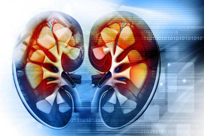
A cross-section graphic of the kidneys.

Chronic kidney disease rarely shows symptoms until the later stages, so screening is recommended for those who are at risk..

Chronic kidney failure, as opposed to acute kidney failure, is a slow and gradually progressive disease. Even if one kidney stops functioning, the other can carry out normal functions. It is not usually until the disease is fairly well advanced and the condition has become severe that signs and symptoms are noticeable; by which time most of the damage is irreversible.

It is important that people who are at high risk of developing kidney disease have their kidney functions regularly checked. Early detection can significantly help prevent serious kidney damage.

The most common signs and symptoms of chronic kidney disease include:

  • anemia
  • blood in urine
  • dark urine
  • decreased mental alertness
  • decreased urine output
  • edema – swollen feet, hands, and ankles (face if edema is severe)
  • fatigue (tiredness)
  • hypertension (high blood pressure)
  • insomnia
  • itchy skin, can become persistent
  • loss of appetite
  • male inability to get or maintain an erection (erectile dysfunction)
  • more frequent urination, especially at night
  • muscle cramps
  • muscle twitches
  • nausea
  • pain on the side or mid to lower back
  • panting (shortness of breath)
  • protein in urine
  • sudden change in bodyweight
  • unexplained headaches

Stages

Changes in the GFR rate can assess how advanced the kidney disease is. In the UK, and many other countries, kidney disease stages are classified as follows:

Stage 1 – GFR rate is normal. However, evidence of kidney disease has been detected.

Stage 2 – GFR rate is lower than 90 milliliters, and evidence of kidney disease has been detected.

Stage 3 – GFR rate is lower than 60 milliliters, regardless of whether evidence of kidney disease has been detected.

Stage 4 – GRF rate is lower than 30 milliliters, regardless of whether evidence of kidney disease has been detected.

Stage 5 – GFR rate is lower than 15 milliliters. Renal failure has occurred.

The majority of patients with chronic kidney disease rarely progress beyond Stage 2. It is important for kidney disease to be diagnosed and treated early for serious damage to be prevented.

Patients with diabetes should have an annual test, which measures microalbuminuria (small amounts of protein) in urine. This test can detect early diabetic nephropathy (early kidney damage linked to diabetes).

Treatment

There is no current cure for chronic kidney disease. However, some therapies can help control the signs and symptoms, reduce the risk of complications, and slow the progression of the disease.

Patients with chronic kidney disease typically need to take a large number of medications. Treatments include:

Anemia treatment

Hemoglobin is the substance in red blood cells that carries vital oxygen around the body. If hemoglobin levels are low, the patient has anemia.

Some kidney disease patients with anemia will require blood transfusions. A patient with kidney disease will usually have to take iron supplements, either in the form of daily ferrous sulfate tablets, or occasionally in the form of injections.

Phosphate balance

People with kidney disease may not be able to eliminate phosphate from their body properly. Patients will be advised to reduce their nutritional phosphate intake – this usually means reducing consumption of dairy products, red meat, eggs, and fish.

High blood pressure

High blood pressure is a common problem for patients with chronic kidney disease. It is important to bring the blood pressure down to protect the kidneys, and subsequently slow down the progression of the disease.

Skin itching

Antihistamines, such as chlorphenamine, may help alleviate symptoms of itching.

Anti-sickness medications

If toxins build up in the body because the kidneys don’t work properly, patients may feel sick (nausea). Medications such as cyclizine or metaclopramide help relieve sickness.

NSAIDs (nonsteroidal anti-inflammatory drugs)

NSAIDs, such as aspirin or ibuprofen should be avoided and only taken if a doctor recommends them.

End-stage treatment

This is when the kidneys are functioning at less than 10-15 percent of normal capacity. Measures used so far – diet, medications, and treatments controlling underlying causes – are no longer enough. The kidneys of patients with end-stage kidney disease cannot keep up with the waste and fluid elimination process on their own – the patient will need dialysis or a kidney transplant in order to survive.

Most doctors will try to delay the need for dialysis or a kidney transplant for as long as possible because they carry the risk of potentially serious complications.

Kidney dialysis

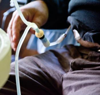
A man undergoing peritoneal dialysis treatment.

Peritoneal dialysis is a treatment option for chronic kidney disease.

This is the removal of waste products and excessive fluids from blood when the kidneys cannot do the job properly any more. Dialysis has some serious risks, including infection.There are two main types of kidney dialysis. Each type also has subtypes. The two main types are:

Hemodialysis: Blood is pumped out of the patient’s body and goes through a dialyzer (an artificial kidney). The patient undergoes hemodialysis about three times per week. Each session lasts for at least 3 hours.

Experts now recognize that more frequent sessions result in a better quality of life for the patient, but modern home-use dialysis machines are making this more regular use of hemodialysis possible.

Peritoneal dialysis: The blood is filtered in the patient’s own abdomen; in the peritoneal cavity which contains a vast network of tiny blood vessels. A catheter is implanted into the abdomen, into which a dialysis solution is infused and drained out for as long as is necessary to remove waste and excess fluid.

Kidney transplant

A kidney transplant is a better option than dialysis for patients who have no other conditions apart from kidney failure. Even so, candidates for kidney transplant will have to undergo dialysis until they receive a new kidney.The kidney donor and recipient should have the same blood type, cell-surface proteins and antibodies, in order to minimize the risk of rejection of the new kidney. Siblings or very close relatives are usually the best types of donors. If a living donor is not possible, the search will begin for a cadaver donor (dead person).

Diet

Following a proper diet is vital for effective kidney failure treatment. Restricting the amount of protein in the diet may help slow down the progression of the disease.

Diet may also help alleviate symptoms of nausea.

Salt intake must be carefully regulated to control hypertension. Potassium and phosphorus consumption, over time, may also need to be restricted.

Vitamin D

Patients with kidney disease typically have low levels of vitamin DVitamin D is essential for healthy bones. The vitamin D we obtain from the sun or food has to be activated by the kidneys before the body can use it. Patients may be given alfacalcidol, or calcitriol.

Fluid retention

People with chronic kidney disease need to be careful with their fluid intake. Most patients will be asked to restrict their fluid intake. If the kidneys do not work properly, the patient is much more susceptible to fluid build-up.

Causes

Kidneys carry out the complex system of filtration in our bodies – excess waste and fluid material are removed from the blood and excreted from the body.

In most cases, kidneys can eliminate most waste materials that our body produces. However, if the blood flow to the kidneys is affected, they are not working properly because of damage or disease, or if urine outflow is obstructed, problems can occur.

In most cases, progressive kidney damage is the result of a chronic disease (a long-term disease), such as:

  • Diabetes – chronic kidney disease is linked to diabetes types 1 and 2. If the patient’s diabetes is not well controlled, excess sugar (glucose) can accumulate in the blood. Kidney disease is not common during the first 10 years of diabetes; it more commonly occurs 15-25 years after diagnosis of diabetes.
  • Hypertension (high blood pressure) – high blood pressure can damage the glomeruli – parts of the kidney involved in filtering waste products.
  • Obstructed urine flow – if urine flow is blocked it can back up into the kidney from the bladder (vesicoureteral reflux). Blocked urine flow increases pressure on the kidneys and undermines their function. Possible causes include an enlarged prostate, kidney stones, or a tumor.
  • Kidney diseases – including polycystic kidney disease, pyelonephritis, or glomerulonephritis.
  • Kidney artery stenosis – the renal artery narrows or is blocked before it enters the kidney.
  • Certain toxins – including fuels, solvents (such as carbon tetrachloride), and lead (and lead-based paint, pipes, and soldering materials). Even some types of jewelry have toxins, which can lead to chronic kidney failure.
  • Fetal developmental problem – if the kidneys do not develop properly in the unborn baby while it is developing in the womb.
  • Systemic lupus erythematosus – an autoimmune disease. The body’s own immune system attacks the kidneys as though they were foreign tissue.
  • Malaria and yellow fever – known to cause impaired kidney function.
  • Some medications – overuse of, for example, NSAIDs (non-steroidal anti-inflammatory drugs), such as aspirin or ibuprofen.
  • Illegal substance abuse – such as heroin or cocaine.
  • Injury – a sharp blow or physical injury to the kidney(s).

Risk factors

The following conditions or situations are linked to a higher risk of developing kidney disease:

  • a family history of kidney disease
  • age – chronic kidney disease is much more common among people over 60
  • atherosclerosis
  • bladder obstruction
  • chronic glomerulonephritis
  • congenital kidney disease (kidney disease which is present at birth)
  • diabetes – one of the most common risk factors
  • hypertension
  • lupus erythematosus
  • overexposure to some toxins
  • sickle cell disease
  • some medications

Diagnosis

A doctor will check for signs and ask the patient about symptoms. The following tests may also be ordered:

  • Blood test – a blood test may be ordered to determine whether waste substances are being adequately filtered out. If levels of urea and creatinine are persistently high, the doctor will most likely diagnose end-stage kidney disease.
  • Urine test – a urine test helps find out whether there is either blood or protein in the urine.
  • Kidney scans – kidney scans may include a magnetic resonance imaging (MRI) scan, computed tomography (CT) scan, or an ultrasound scan. The aim is to determine whether there are any blockages in the urine flow. These scans can also reveal the size and shape of the kidneys – in advanced stages of kidney disease the kidneys are smaller and have an uneven shape.
  • Kidney biopsy – a small sample of kidney tissue is extracted and examined for cell damage. An analysis of kidney tissue makes it easier to make a precise diagnosis of kidney disease.
  • Chest X-ray – the aim here is to check for pulmonary edema (fluid retained in the lungs).
  • Glomerular filtration rate (GFR) – GFR is a test that measures the glomerular filtration rate – it compares the levels of waste products in the patient’s blood and urine. GFR measures how many milliliters of waste the kidneys can filter per minute. The kidneys of healthy individuals can typically filter over 90 ml per minute.

Complications

If the chronic kidney disease progresses to kidney failure, the following complications are possible:

  • anemia
  • central nervous system damage
  • dry skin or skin color changes
  • fluid retention
  • hyperkalemia, when blood potassium levels rise, possibly resulting in heart damage
  • insomnia
  • lower sex drive
  • male erectile dysfunction
  • osteomalacia, when bones become weak and break easily
  • pericarditis, when the sac-like membrane around the heart becomes inflamed
  • stomach ulcers
  • weak immune system

Prevention

Managing the chronic condition

Some conditions increase the risk of chronic kidney disease (such as diabetes). Controlling the condition can significantly reduce the chances of developing kidney failure. Individuals should follow their doctor’s instructions, advice, and recommendations.

Diet

A healthy diet, including plenty of fruits and vegetables, whole grains, and lean meats or fish will help keep blood pressure down.

Physical activity

Regular physical exercise is ideal for maintaining healthy blood pressure levels; it also helps control chronic conditions such as diabetes and heart disease. Individuals should check with a doctor that an exercise program is suited to their age, weight, and health.

Avoiding certain substances

Including abusing alcohol and drugs. Avoid long-term exposure to heavy metals, such as lead. Avoid long-term exposure to fuels, solvents, and other toxic chemicals.


MedicalNewsToday

#Everything you need to know about gout

Postado em

Gout is a common type of arthritis that causes intense pain, swelling, and stiffness in a joint. It usually affects the joint in the big toe.

Gout attacks can come on quickly and keep returning over time, slowly harming tissues in the region of the inflammation, and can be extremely painful. Hypertension, cardiovascular, and obesityare risk factors for gout.

It is the most common form of inflammatory arthritis in men, and although it is more likely to affect men, women become more susceptible to it after the menopause.

The Centers for Disease Control and Prevention (CDC) report that 8.3 million Americans were affected by gout between 2007 to 2008.

Fast facts on goutHere are some key points about gout. More detail and supporting information is in the main article.

  • Gout is a form of arthritis caused by excess uric acid in the bloodstream.
  • The symptoms of gout are due to the formation of uric acid crystals in the joints and the body’s response to them.
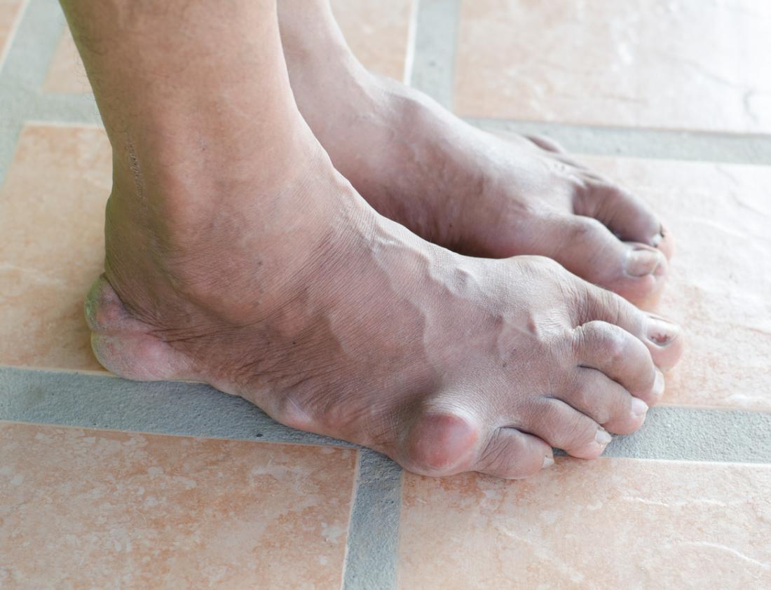
  • Gout most classically affects the joint in the base of the big toe.
  • Gout attacks often occur without warning in the middle of the night.
  • Most gout cases are treated with specific medications.

Treatment

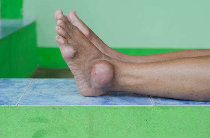
person with gout

Gout patients often have acute inflammation around their joints.

The majority of gout cases are treated with medication. Medication can be used to treat the symptoms of gout attacks, prevent future flares, and reduce the risk of gout complications such as kidney stones and the development of tophi.

Commonly used medications include nonsteroidal anti-inflammatory drugs (NSAIDs), colchicine, or corticosteroids. These reduce inflammation and pain in the areas affected by gout and are usually taken orally.

Medications can also be used to either reduce the production of uric acid (xanthine oxidase inhibitors such as allopurinol) or improve the kidney’s ability to remove uric acid from the body (probenecid).

Without treatment, an acute gout attack will be at its worst between 12 and 24 hours after it began. A person can expect to recover within 1 to 2 weeks without treatment, but there may be significant pain during this period.

Tests and diagnosis

Gout can be tricky to diagnose, as its symptoms, when they do appear, are similar to those of other conditions. While hyperuricemia occurs in the majority of people that develop gout, it may not be present during a flare. On top of that, the majority of people with hyperuricemia do not develop gout.

One diagnostic test that doctors can carry out is the joint fluid test, where fluid is extracted from the affected joint with a needle. The fluid is then examined to see if any urate crystals are present.

As joint infections can also cause similar symptoms to gout, a doctor can look for bacteria when carrying out a joint fluid test in order to rule a bacterial cause. The fluid may need to be sent to a lab, where it can take several days to analyze.

Doctors can also do a blood test to measure the levels of uric acid in the blood, but, as mentioned, people with high uric acid levels do not always experience gout. Equally, some people can develop the symptoms of gout without having increased levels of uric acid in the blood.

Finally, doctors can search for urate crystals around joints or within a tophus using ultrasound scan. X-rays cannot detect gout, but may be used to rule out other causes.

Types

There are various stages through which gout progresses, and these are sometimes referred to as different types of gout.

Asymptomatic hyperuricemia

It is possible for a person to have elevated uric acid levels without any outward symptoms. At this stage, treatment is not required, though urate crystals may deposit in tissue and cause slight damage.

People with asymptomatic hyperuricemia may be advised to take steps to address any possible factors contributing to uric acid build-up.

Acute gout

This stage occurs when the urate crystals that have been deposited suddenly cause acute inflammation and intense pain. This sudden attack is referred to as a “flare” and will normally subside within 3 to 10 days. Flares can sometimes be triggered by stressful events, alcohol and drugs, as well as cold weather.

Interval or intercritical gout

This stage is the period in between attacks of acute gout. Subsequent flares may not occur for months or years, though if not treated, over time, they can last longer and occur more frequently. During this interval, further urate crystals are being deposited in tissue.

Chronic tophaceous gout

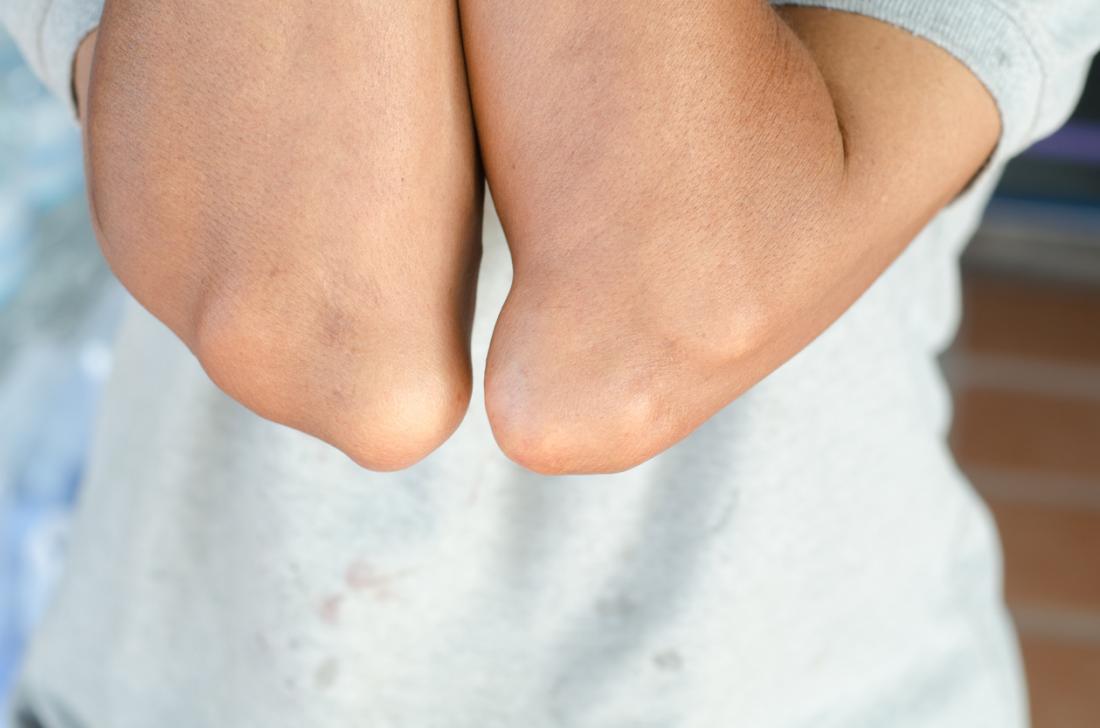
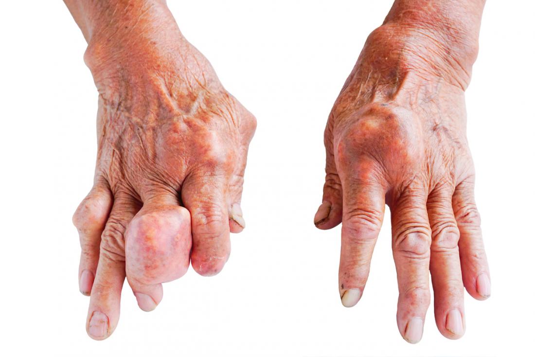
Chronic tophaceous gout is the most debilitating type of gout. Permanent damage may have occurred in the joints and the kidneys. The patient can suffer from chronic arthritis and develop tophi, big lumps of urate crystals, in cooler areas of the body such as the joints of the fingers.

It takes a long time without treatment to reach the stage of chronic tophaceous gout – around 10 years. It is very unlikely that a patient receiving proper treatment would progress to this stage.

Pseudogout

One condition that is easily confused with gout is pseudogout. The symptoms of pseudogout are very similar to those of gout, although thr flare-ups are usually less severe.

The major difference between gout and pseudogout is that the joints are irritated by calciumpyrophosphate crystals rather than urate crystals. Pseudogout requires different treatment to gout.

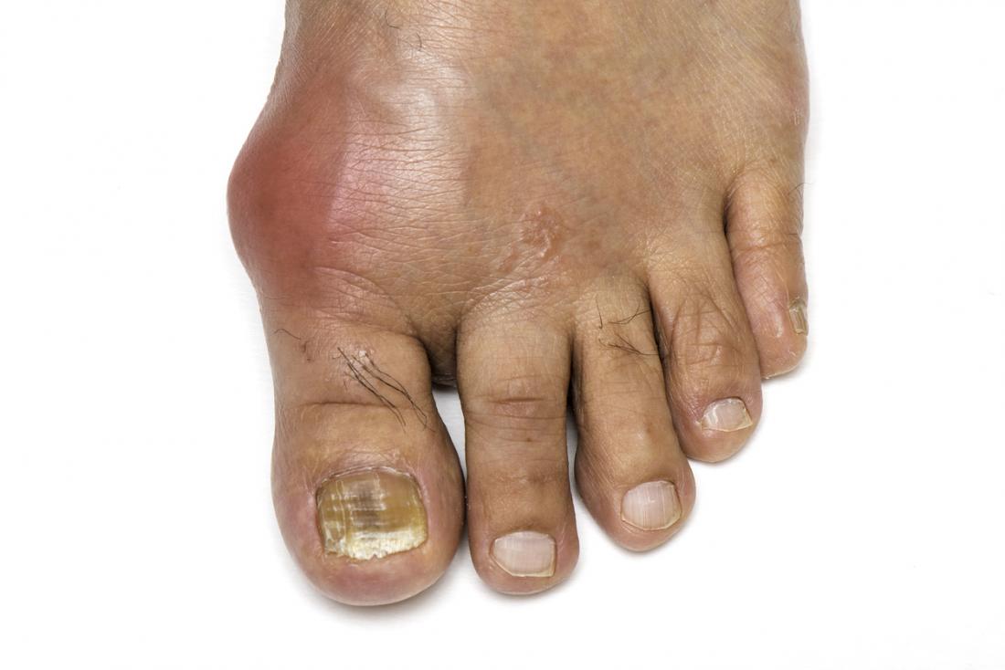
Pictures

The following images show the appearance of gout when it develops.

1of4

Causes

Gout is caused initially by an excess of uric acid in the blood, or hyperuricemia. Uric acid is produced in the body during the breakdown of purines – chemical compounds that are found in high amounts in certain foods such as meat, poultry, and seafood.

Normally, uric acid is dissolved in the blood and is excreted from the body in urine via the kidneys. If too much uric acid is produced, or not enough is excreted, it can build up and form needle-like crystals that trigger inflammation and pain in the joints and surrounding tissue.

Risk factors

There are a number of factors that can increase the likelihood of hyperuricemia, and therefore gout:

Age and gender: Men produce more uric acid than women, though women’s levels of uric acid approach those of men after the menopause.

Genetics: A family history of gout increases the likelihood of the condition developing.

Lifestyle choices: Alcohol consumption interferes with the removal of uric acid from the body. Eating a high-purine diet also increases the amount of uric acid in the body.

Lead exposure: Chronic lead exposure has been linked to some cases of gout.

Medications: Certain medications can increase the levels of uric acid in the body; these include some diuretics and drugs containing salicylate.

Weight: Being overweight increases the risk of gout as there is more turnover of body tissue, which means more production of uric acid as a metabolic waste product. Higher levels of body fat also increase levels of systemic inflammation as fat cells produce pro-inflammatory cytokines.

Recent trauma or surgery: Increases risk.

Other health problems: Renal insufficiency and other kidney problems can reduce the body’s ability to efficiently remove waste products, leading to elevated uric acid levels. Other conditions associated with gout include high blood pressure and diabetes.

Symptoms

Gout usually becomes symptomatic suddenly without warning, often in the middle of the night.

The main symptoms are intense joint pain that subsides to discomfort, inflammation, and redness.

Gout frequently affects the large joint of the big toe, but can also affect the forefoot, ankles, knees, elbows, wrists, and fingers.

The pain can be excruciating. A veteran visiting a Hospital in Birmingham, AL, said:

“I’ve been shot, beat up, stabbed, and thrown out of a helicopter, but none of that compared to the gout.”

Complications

In some cases, gout can develop into more serious conditions, such as:

  • Kidney stones: If urate crystals collect in the urinary tract, they can become kidney stones.
  • Recurrent gout: Some people only ever have one flare up; others may have regular recurrences, causing gradual damage to the joints and surrounding tissue.

Prevention tips

There are many lifestyle and dietary guidelines that can be tried to protect against flares or prevent gout from occurring in the first instance:

  • maintain a high fluid intake of around 2 to 4 liters a day
  • avoid alcohol
  • maintain a healthy body weight

Home remedies

Individuals with gout can manage flare-ups by moderating their diet. A balanced diet can help reduce symptoms.

Decreasing foods that are high in purines, to ensure that the levels of uric acid in the blood do not get too high, is reasonable to try. Here is a list of high-purine foods to be wary of:

  • anchovies
  • asparagus
  • beef kidneys
  • brains
  • dried beans and peas
  • game meats
  • gravy
  • herring
  • liver
  • mackerel
  • mushrooms
  • sardines
  • scallops
  • sweetbreads

While it is reasonable to decrease or avoid these foods, it has been found that a high purine-rich diet does not increase the risk of gout, or aggrevate symptoms in research studies.

Asparagus, beans, some other plant-based foods, and mushrooms are also sources of purines, but research suggests that these do not trigger gout attacks and do not impact uric acid levels.

Various epidemiological studies have shown that purine-rich vegetables, whole grains, nuts and legumes, and less sugary fruits, coffee, and vitamin C supplements lower blood uric acid levels, but do not decrease the risk of gout. Red meat, fructose-containing beverages, and alcohol can increase the risk.

The role of uric acid in gout has been clearly defined and understood. As a result of this and the wide availability of relevant medications, gout is a very controllable form of arthritis.

MedicalNewsToday

#Kidney disease: Does gout increase risk?

Postado em Atualizado em

Building on previous findings, a new study concludes that people with gout are more likely to develop kidney disease. The authors believe that the results offer new ways to reduce the risk of chronic kidney disease.
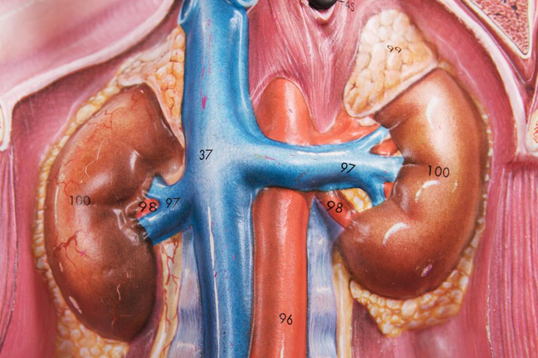
Kidney anatomy model

A recent study looks at whether gout influences kidney health.

Chronic kidney disease (CKD) is a long term condition, characterized by reduced kidney function.

CKD impacts an estimated 14% of people in the United States.

Researchers have already pinned down a number of risk factors for CKD, including high blood pressurediabetes, and smoking.

However, as the authors of the most recent study explain, “There is increasing interest by the scientific community in the role of novel risk factors.”

The latest research, published in BMJ Open, investigates the potential role of gout as a risk factor for CKD.

Gout is both surprisingly common and surprisingly painful. It is caused by elevated levels of uric acid in the blood, which is referred to as hyperuricemia.

Over time, uric acid can build up, causing sharp crystals to form in joints, most commonly of the big toe.

Gout, a form of inflammatory arthritis, affects more than 8.3 million people in the U.S.

Gout and the kidney

Earlier studies have identified links between gout and kidney function. For instance, one paper, published in 2012, found that poor kidney function was a risk factor for gout.

Other studies, in animal models, have shown that increased uric acid in the blood has the potential to damage the kidneys.

Previous research has also highlighted a relationship between raised uric acid levels and kidney function. For instance, one study involving people with hyperuricemia found that taking drugs that reduce levels of uric acid also reduced the severity of kidney dysfunction.

Because both gout and CKD are common, understanding how the two conditions interact is important; and despite previous work, no studies have conclusively shown that gout contributes to kidney failure.

To investigate, the researchers drew information from 68,897 adults aged 18 or older with gout in the United Kingdom and compared it with data from 554,964 matched individuals without gout. They followed both groups for an average of 3.68 years.

For the study, they defined advanced kidney disease as the first occurrence of:

  • dialysis, transplant, or end stage kidney disease
  • kidney function at less than 10% of normal
  • death associated with CKD
  • a doubling of serum creatinine from the baseline levels

The medical community considers creatinine to be a reliable marker of kidney function.

‘Astonishing’ findings

As part of the analysis, the scientists controlled for several factors that might otherwise influence the results, including high blood pressure, diabetes, heart disease, drugs that impact kidney function, smoking status, and alcohol use.

Even after accounting for these factors, the results were significant.

“While we always believed that high levels of uric acid might be bad for kidneys and that patients with gout may have a higher risk of kidney failure, we were quite surprised by the magnitude of the risk imposed by gout in these patients,” explains lead author Prof. Austin Stack from the University of Limerick, in Ireland.

The results were quite astonishing. […] We discovered that patients who suffered from gout had a 29% higher risk of advanced CKD, compared to those without gout.”

Prof. Austin Stack

The strongest statistical relationship occurred among individuals with end stage kidney disease, as the authors explain:

“The magnitude of the gout–CKD association was greatest for a diagnosis of [end stage kidney disease], with over a twofold higher risk.”

According to Prof. Stack, “Taken together, the findings from this study suggest that gout is an independent risk factor for progression of CKD and kidney failure.”

This paper is important and adds a new level to our understanding of CKD. Because CKD is so prevalent, understanding the risk factors involved might provide new ways to control it.

The current study has many strengths, including the collection of a vast amount of data. However, the authors do outline certain limitations.

For instance, the authors could only assess whether someone had gout by observing doctors’ diagnoses or registering their use of urate lowering treatments. In reality, gout often goes undiagnosed, so the analysis likely missed some individuals.

Although the scientists accounted for a wide range of factors in their analysis, because the study is observational, there is the possibility that an unmeasured variable plays a pivotal role.

Overall, these findings add to the evidence that uric acid, gout, and kidney health are linked. The authors hope that future work can ascertain whether “adequate control of gout can reduce the risk of CKD progression.”

MedicalNewsToday

#Considerações sobre cuidados paliativos pediátricos

Postado em

médica atendendo criança no hospital

Considerações sobre cuidados paliativos pediátricos


A Organização Mundial de Saúde (OMS) define cuidados paliativos como a prevenção e o alívio do sofrimento de pacientes adultos e pediátricos e de suas famílias, que enfrentam os problemas associados a doenças potencialmente fatais. Esses problemas incluem o sofrimento físico, psicológico, social e espiritual dos pacientes e de seus familiares.

Para a OMS, os cuidados paliativos:

  • Proporcionam alívio da dor e de outros sintomas angustiantes;
  • Afirmam a vida e consideram a morte como um processo normal;
  • Não pretendem apressar ou adiar a morte;
  • Integram os aspectos psicológicos e espirituais do atendimento ao paciente;
  • Oferecem um sistema de apoio para ajudar os pacientes a viver o mais ativamente possível até a morte e para auxiliar a família durante a doença do paciente e em seu próprio luto;
  • Usam uma abordagem de equipe para atender às necessidades dos pacientes e de suas famílias, incluindo o aconselhamento do luto, se indicado;
  • Melhoram a qualidade de vida, e também podem influenciar positivamente o curso da doença;
  • São aplicáveis no início do curso da doença, em conjunto com outras terapias que se destinam a prolongar a vida, como a quimioterapia ou a radioterapia, e incluem as investigações necessárias para melhor compreender e gerir complicações clínicas angustiantes.

Cuidados Paliativos em Pediatria

Os cuidados paliativos em pediatria (CPP) evoluíram do movimento de cuidados paliativos para adultos, estabelecido nas décadas de 1970 e 19803. Representam um campo especial, embora intimamente relacionado aos cuidados paliativos de adultos (CPA). Os CCP, segundo a OMS, são definidos como o cuidado ativo total do corpo, mente e espírito da criança, e isso envolve também dar apoio à família. De acordo com a OMS:

  • CPP começam quando a doença é diagnosticada e continuam independentemente de a criança receber ou não tratamento direcionado à doença;
  • Os profissionais de saúde devem avaliar e aliviar o sofrimento físico, psicológico e social de uma criança;
  • CPP eficazes requerem uma ampla abordagem multidisciplinar que inclua a família e faça uso dos recursos disponíveis na comunidade; podem ser implementados com sucesso mesmo que os recursos sejam limitados.
  • Podem ser fornecidos em instalações de cuidados terciários, em centros de saúde comunitários e até em lares de crianças.

Para os profissionais que trabalham em pediatria, é incontestável que uma criança não é simplesmente um adulto em miniatura. Isto é bastante verdadeiro em CPP, já que os cuidadores devem se esforçar para abordar os aspectos mais amplos das necessidades psicossociais e educacionais de uma criança. No entanto, os conhecimentos alcançados em cuidados paliativos para adultos são extremamente relevantes e devem ser bem aproveitados, a começar pela vantagem inicial de 40 ou 50 anos na frente.

Os princípios da medicina paliativa incluem uma abordagem holística focada no paciente, desenvolvimento e confiança em evidências robustas para tomar decisões terapêuticas racionais. E uma ênfase constante na combinação desses dois fatores para equilibrar a carga e os benefícios, assegurando que os melhores interesses do indivíduo permaneçam preservados. Estes tornam-se particularmente importantes em cuidados paliativos, quando o objetivo do tratamento se torna a manutenção da qualidade de vida.

Algumas similaridades e diferenças entre CPA e CPP estão descritas nos Quadros 1 e 2.

Indicações

Os CPP são idealmente integrados ao cuidado de qualquer criança com uma condição potencialmente fatal e são indicados e observados nas seguintes condições citadas nos Quadros 3 e 4.

Dor e outros sintomas em CPP

As crianças que necessitam de CPP apresentam uma série de manifestações clínicas. As mais comuns, independentemente do diagnóstico, são dordispneia e fadiga. Em cuidados paliativos, há uma forte relação entre componentes físicos e psicológicos das diversas manifestações clínicas. Distúrbios do sono, ansiedade, tristeza e depressão podem ter impacto significativo na dor. A dor e o sofrimento repercutem não apenas no paciente, mas também em seus familiares.

O Quadro 5 descreve as manifestações clínicas mais comuns de algumas situações que exigem cuidados paliativos em pediatria

Apesar do crescimento e dos avanços significativos dos CPP nas últimas duas décadas, atualmente um grande número de hospitais infantis não possui um serviço específico ou os recursos são insuficientes.

Para Friedrichsdorf e Bruera (2018), a implementação de um serviço de CPP passa em geral por quatro etapas. Isso deve ser antecipado, e um plano de ação pode incluir:

  • Negação: documentar as necessidades não atendidas, realizar pesquisas entre funcionários, pacientes e provedores;
  • Palliphobia: estreita colaboração com colegas; planejamento disciplinado, rápida resolução de conflitos;
  • Pallilalia: documentação do valor dos CPP para a liderança; grandes rodadas por especialistas em campo e revisão externa;
  • Paliativo: “faça o bem e fale sobre isso” – inovadores clínicos e administrativos devem estar envolvidos no desenvolvimento do programa;
  • Financiamento seguro: “verifique se sua paixão está conectada ao sistema de pagamento de alguém”.

Os CPP de alta qualidade para crianças com doenças graves agora são um padrão esperado da medicina. No entanto, mesmo em ambientes ricos em recursos, ainda existem barreiras significativas para alcançar atendimento ideal relacionado à falta de educação formal, questões de reembolso, impacto emocional ao se cuidar de uma criança moribunda e, o mais importante, falta de equipes interdisciplinares de CPP.

O tratamento deve se concentrar, sempre que possível, nos esforços contínuos para controlar a doença subjacente. Ao mesmo tempo, as crianças e suas famílias devem ter acesso a cuidados interdisciplinares visando a promover o bem-estar físico, psicológico e espiritual ideal. Mitos e conceitos errôneos persistentes levam ao controle inadequado dos sintomas em crianças com doenças que limitam a vida.

Para Friedrichsdorf e Bruera (2018), os CPP defendem a prestação de cuidados de conforto, dor e gerenciamento de sintomas simultaneamente com os tratamentos direcionados à doença.


PebMed

Referências:

#Novo antibiótico aprovado para tratamento de pneumonia comunitária nos EUA

Postado em Atualizado em

antibioticos

Novo antibiótico aprovado para tratamento de pneumonia comunitária nos EUA


No dia 19 de agosto, a U.S. Food and Drug Administration (FDA) aprovou um novo antibiótico para o tratamento de pneumonia comunitária: a lefamulina.

Com o nome comercial de Xenleta®, a lefamulina é um antimicrobiano que inibe a síntese proteica bacteriana ao interagir com a subunidade 50s do rRNA. Tem atividade bactericida contra cepas de Streptococcus pneumoniaeHaemophilus influenzaeChlamydophila pneumoniaeLegionella pneumophila e Mycoplasma pneumoniae, e bacteriostática contra Staphylococcus aureus sensível à meticilina e Streptococcus pyogenes in vitro. Não é ativa contra enterobactérias ou Pseudomonas aeruginosa e não tem comprovação de eficácia em infecções causadas por MRSA.

O novo antibiótico foi aprovado após 2 ensaios clínicos multicêntricos, duplo-cegos e randomizados. Compreendeu 1289 pacientes em que lefamulina por 5-10 ou 7 dias foi não-inferior ao tratamento com moxifloxacino com ou sem linezolida por 7-10 dias. Ou 7 dias de moxifloxacino em pacientes com quadro de pneumonia comunitária bacteriana.

Durante os estudos, os eventos adversos mais frequentemente reportados consistiram em reações no local de infusão, diarreia, náuseas, vômitos, elevação de enzimas hepáticas, hipocalemia, insônia e cefaleia. Além disso, o uso de lefamulina tem o potencial de prolongar o intervalo QT. E, por causa disso, deve ser evitado em pacientes com prolongamento de intervalo QT conhecido, pacientes com arritmias ventriculares, incluindo torsade de pointes, pacientes em uso de antiarrítmicos de classe IA (como quinidina ou procainamida) ou de classe III (como amiodarona e sotalol), e pacientes em uso de outras medicações com potencial de prolongar o intervalo QT.

Xenleta® está disponível nas formulações intravenosa e oral e foi aprovado com as posologias de 150 mg, EV, 12/12 horas ou 600 mg, VO, 12/12 horas. Não há necessidade de ajuste na disfunção renal, porém pacientes com disfunção hepática grave (Child C) devem ter a dose reduzida para 150 mg, EV, a cada 24 horas.

A lefamulina ainda não tem previsão de chegar ao Brasil, mas constitui uma nova opção para o tratamento de pneumonia comunitária.


PebMed


Autora: 

Referências:

#Gravidez: álcool danifica a placenta mesmo na altura da conceção

Postado em

Fonte de imagem: Medical News Today

O consumo de bebidas alcoólicas durante a gravidez foi associado, num estudo, a um fraco desenvolvimento da placenta, podendo causar problemas como restrição no crescimento fetal e baixo peso à nascença.

O estudo conduzido pelas investigadoras Jacinta Kalisch-Smith e Karen Moritz da Universidade de Queensland na Austrália analisou o impacto do consumo de álcool sobre a placenta e sobre o desenvolvimento do embrião no estado inicial da gravidez.

Para o efeito, as investigadoras usaram ratazanas que foram expostas a álcool na altura da conceção.

“Através de um modelo de ratazanas, avaliámos a capacidade de o embrião se implantar no útero e, mais tarde, a formação dos vasos sanguíneos na placenta”, explicou Jacinta Kalisch-Smith.

Assim, a equipa conseguiu estudar as alterações no decorrer da gestação e descobriram que a exposição ao álcool, mesmo que precoce (entre quatro dias antes e quatro dias após a fertilização), restringia o crescimento e função da placenta.

“Descobrimos que a exposição inicial ao álcool reduzia a formação de vasos sanguíneos na placenta, e que isso fez com que menos nutrientes fossem fornecidos ao embrião”, divulgou Jacinta Kalisch-Smith.

Curiosamente, as placentas de embriões femininos demonstraram uma maior suscetibilidade, com uma redução de até 17% no seu tamanho e de 32% na formação de vasos sanguíneos, limitando a capacidade de transporte de nutrientes pela placenta.

Jacinta Kalisch-Smith explicou que estes resultados são relevantes para a saúde humana pois ajudam a explicar em parte a razão pela qual os bebés expostos a álcool dentro do útero frequentemente nascem com peso baixo, um fator de risco para a diabetes de tipo 2, hipertensão e obesidade na idade adulta.

#Fármaco atrasa diabetes de tipo 1 em pacientes de alto risco

Postado em Atualizado em

Fonte de imagem: Reach Farther

Um tratamento com um fármaco de imunoterapia conseguiu desacelerar a progressão da diabetes de tipo 1 num ensaio que contou com pessoas de alto risco.

Conhecido como teplizumab, o fármaco de imunoterapia consiste num anticorpo monoclonal anti-CD3 e demonstrou evitar o desencadeamento da doença autoimune em pelo menos dois anos.

O ensaio clínico, denominado Type 1 Diabetes TrialNet, foi uma colaboração internacional com o objetivo de identificar formas de adiar ou evitar a diabetes de tipo 1.

Para o ensaio, uma equipa de investigadores recrutou 76 participantes com idades compreendidas entre os oito e os 49 anos. Os participantes eram familiares de pacientes com diabetes de tipo 1 e apresentavam pelo menos dois tipos de autoanticorpos relacionados com a diabetes e uma tolerância anormal à glicose.

Os participantes foram divididos em dois grupos, tendo um dos grupos recebido teplizumab durante 14 dias, e o outro grupo um placebo.

Todos os participantes foram submetidos a testes regulares de tolerância à glicose até ao final do estudo, ou até desenvolverem diabetes de tipo 1 clínica, se esta sucedesse primeiro.

No decorrer do ensaio, 72% dos participantes do grupo de controlo desenvolveram diabetes clínica, contra 43% do grupo do teplizumab.

A mediana de tempo para os participantes do grupo de controlo desenvolverem diabetes clínica foi de um pouco mais de 24 meses, enquanto no grupo de tratamento a mediana de tempo foi de 48 meses.

Os efeitos do fármaco foram mais pronunciados durante o primeiro ano após o tratamento. Durante esse período, 41% dos participantes desenvolveram diabetes clínica, maioritariamente no grupo de controlo.

“A diferença nos resultados foi incrível. Esta descoberta é a primeira evidência que vimos em como a diabetes de tipo 1 clínica pode ser atrasada com um tratamento preventivo precoce”, comentou Lisa Spain, cientista neste projeto, dos Institutos Nacionais de Saúde dos EUA.

#Vitamina K insuficiente associada a incapacidade em idosos

Postado em

Fonte de imagem: BodyNutrition

Um estudo recente apurou que os adultos de idade mais avançada com níveis reduzidos de vitamina K circulante poderão apresentar um maior risco de limitações e de incapacidade de mobilidade.

A vitamina K circulante reflete a quantidade dessa vitamina na alimentação seguida. As melhores fontes de vitamina K são as folhosas verdes como o espinafre, os brócolos e a couve kale, e ainda alguns produtos láteos. Uma chávena de espinafre cru oferece 181% da dose diária recomendada de vitamina K a um adulto e metade de uma chávena de brócolos cozidos contém 138%.

Para o estudo, que foi liderado por investigadores da Universidade Tuffs, EUA, foram analisados dados de 635 homens e 688 mulheres com idades compreendidas entre os 70 e os 79 anos de idade.

A mobilidade dos participantes foi avaliada a cada seis meses, durante um período de seis a 10 anos, através de consultas clínicas e entrevistas por via telefónica.

Os investigadores consideraram como limitações na mobilidade dois relatos semestrais, consecutivos, de qualquer nível de dificuldade em caminhar 400 metros ou subir 10 degraus sem descansar.

A incapacidade de mobilidade foi definida como dois relatos semestrais consecutivos de muita dificuldade ou incapacidade de caminhar a mesma distância ou subir a mesma quantidade.

Foi apurado que os adultos mais velhos com níveis reduzidos de vitamina K circulante (filoquinona) eram quase 1,5 vezes mais propensos a desenvolverem limitações na mobilidade e quase o dobro da possibilidade de desenvolverem incapacidade de mobilidade em comparação com os que registavam níveis suficientes.

“O estado de pouca vitamina K tem sido associado ao desenvolvimento de doenças crónicas que conduzem à incapacidade, mas o trabalho para perceber esta ligação está ainda nos primórdios. Desenvolvemos aqui estudos anteriores que descobriram que os níveis reduzidos de vitamina K estão associados a um ritmo mais lento e um maior risco de osteoartrite”, concluiu Kyla Shea, primeira autora do estudo.